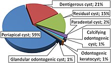
Calcifying odontogenic cyst
[3] It can appear in any location in the oral cavity, but more commonly affects the anterior (front) mandible and maxilla.
On dental radiographs, the calcifying odontogenic cyst appears as a unilocular (one circle) radiolucency (dark area).
[2] They are normally presented as a painless, slow-growing mass on the mandible and/or the maxilla, mostly in the front of the mouth.
When a COC is located in the maxilla, individuals might complain of nasal stiffness, epistaxis, and headache.
Calcifying odontogenic cyst can is the presence of a variable number of ghost cells within the epithelial lining.
[7] The mechanism for the formation of a calcifying odontogenic cyst is controversial, whether the ghost cells change is based on coagulative necrosis (accidental cell death caused by ischemia or infarction)/the build up of enamel protein or it's a form of normal or abnormal keratinization (formation of keratin proteins) of odontogenic epithelium.
It first appears as fine basophilic granules that increase in size and number forming large masses of calcifying material.
Eosinophilic dentinoid (abnormal form of dentin) material is present next to a sheet of ghost cells.
[7] Some forms of the cystic type of COC, the epithelial lining proliferates into the lumen (inside space of the cyst) so its filled with masses of ghost cells and dystrophic calcifications.
[7] In a different form, unifocal or multifocal epithelial proliferation (increase in numbers) of the cyst lining into the lumen may look similar to ameloblastoma.
Intraosseous consists of ameloblastoma-like strands and epithelium in fibrous connective tissue stroma with ghost cells present.
They are used to confirm the presence of calcifications along the cyst wall that were not detected in the radiographic images.
Formation of calcified tissues in the lumen of the cyst wall showing dystrophic calcification.
[8] The standard treatment of calcifying odontogenic cyst is enucleation and curettage, however it depends on the lesion site and histological pattern.
[10] In a case study that was conducted in 2011, a 23-year-old female came with in swelling in the upper right side of the jaw that had been present for about 2 years.
[11] The location of the lesion is important for diagnosis because many other bone diseases can be commonly found in the posterior mandible.