Camillo Golgi
[1] Golgi and the Spanish biologist Santiago Ramón y Cajal were jointly given the Nobel Prize in Physiology or Medicine 1906 "in recognition of their work on the structure of the nervous system".
Three years his junior, Bizzozero was an eloquent teacher and experimenter, who specialised in histology of the nervous system and the properties of bone marrow.
[5] Financial pressure prompted him to join the Hospital of the Chronically Ill (Pio Luogo degli Incurabili) in Abbiategrasso, near Milan, as Chief Medical Officer in 1872.
To continue research, he set up a simple laboratory on his own in a refurbished hospital kitchen, and it was there that he started making his most notable discoveries.
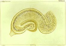
His major achievement was the development of staining technique for nerve tissue called the black reaction (later the Golgi's method).
The silver chromate precipitate, as a reaction product, selectively stains only some cellular components randomly, sparing other cell parts.
The silver chromate particles create a stark black deposit on the soma (nerve cell body) as well as on the axon and all dendrites, providing an exceedingly clear and well-contrasted picture of neuron against a yellow background.
[11] On 16 February 1873, he wrote to his friend Niccolò Manfredi: I am delighted that I have found a new reaction to demonstrate, even to the blind, the structure of the interstitial stroma of the cerebral cortex.His discovery was published in the Gazzeta Medica Italiani on 2 August 1873.
Using his black reaction, Golgi could trace various regions of the cerebro-spinal axis, clearly distinguishing the different nervous projections, namely axon from the dendrites.
Thus, Golgi presented the reticular theory which states that the brain is a single network of nerve fibres, and not of discrete cells.
[13][14] Although Golgi's earlier works between 1873 and 1885 clearly depicted the axonal connections of cerebellar cortex and olfactory bulb as independent of one another, his later works including the Nobel Lecture showed the entire granular layer of the cerebellar cortex occupied by a network of branching and anastomosing nerve processes.
[16] In addition to this, Golgi was the first to give clear descriptions of the structure of the cerebellum, hippocampus, spinal cord, olfactory lobe, as well as striatal and cortical lesions in a case of chorea.
[17] He further developed a stain specific for myelin (a specialised membrane which wraps around the axon) using potassium dichromate and mercuric chloride.
He was the first to dissect out intact nephrons, and show that the distal tubulus (loop of Henle) of the nephron returns to its originating glomerulus, a finding that he published in 1889 ("Annotazioni intorno all'Istologia dei reni dell'uomo e di altri mammifieri e sull'istogenesi dei canalicoli oriniferi".
[18] A French Army physician Charles Louis Alphonse Laveran discovered that malaria was caused by microscopic parasite (now called Plasmodium falciparum) in 1880.
By 1898, along with Giovanni Battista Grassi, Amico Bignami, Giuseppe Bastianelli, Angelo Celli and Marchiafava, he confirmed that malaria was transmitted by anopheline mosquitoes.
[23] He noticed thread-like networks inside the cells and named them apparato reticolare interno (internal reticular apparatus).
[26] Golgi, together with Santiago Ramón y Cajal, received the Nobel Prize in Physiology or Medicine in 1906 for his studies of the structure of the nervous system.