Hippocampus
In Alzheimer's disease (and other forms of dementia), the hippocampus is one of the first regions of the brain to suffer damage; short-term memory loss and disorientation are included among the early symptoms.
Many neurons in the rat and mouse hippocampi respond as place cells: that is, they fire bursts of action potentials when the animal passes through a specific part of its environment.
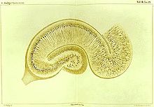
The earliest description of the ridge running along the floor of the inferior horn of the lateral ventricle comes from the Venetian anatomist Julius Caesar Aranzi (1587), who likened it first to a silkworm and then to a seahorse (Latin hippocampus, from Greek ἱππόκαμπος, from ἵππος, 'horse' + κάμπος, 'sea monster').
[14] Many studies later culminating in work by Papez, and MacLean, the involvement of other interacting brain regions associated with emotion was recognised.
[15] The hippocampus is anatomically connected to parts of the brain that are involved with emotional behavior, including the septal area, the hypothalamic mammillary bodies, and the anterior nuclear complex in the thalamus.
[16][17] The hippocampus is an inward fold of three-layered archicortex (one of three regions of the allocortex) into the medial temporal lobe of the brain, where it elevates into the floor of each lateral ventricle inferior horn.
[22] The hippocampal formation refers to the hippocampus, and its related adjoining parts to include the dentate gyrus, the subiculum, the presubiculum, parasubiculum, and the entorhinal cortex.
From there, CA3 axons called Schaffer collaterals leave the deep part of the cell body and loop up to the apical dendrites and then extend to CA1 (third synapse).
There continues to be some interest in hippocampal olfactory responses, in particular, the role of the hippocampus in memory for odors, but few specialists today believe that olfaction is its primary function.
British psychologist Jeffrey Gray developed this line of thought into a full-fledged theory of the role of the hippocampus in anxiety, called the behavioral inhibition system.
Although it had historical precursors, this idea derived its main impetus from a famous report by American neurosurgeon William Beecher Scoville and British-Canadian neuropsychologist Brenda Milner.
Neural activity sampled from 30 to 40 randomly chosen place cells carries enough information to allow a rat's location to be reconstructed with high confidence.
[61] In some cases, the firing rate of hippocampal cells depends not only on place but also the direction a rat is moving, the destination toward which it is traveling, or other task-related variables.
[100] fMRI findings from studies in approach-avoidance decision-making found evidence for a functional role that is not explained by either long-term memory or spatial cognition.
Overall findings showed that the anterior hippocampus is sensitive to conflict, and that it may be part of a larger cortical and subcortical network seen to be important in decision-making in uncertain conditions.
[112] The underlying currents producing the theta wave are generated mainly by densely packed neural layers of the entorhinal cortex, CA3, and the dendrites of pyramidal cells.
These reflect subthreshold membrane potentials and strongly modulate the spiking of hippocampal neurons and synchronise across the hippocampus in a travelling wave pattern.
[123] It has been proposed that sharp waves are, in fact, reactivations of neural activity patterns that were memorized during behavior, driven by strengthening of synaptic connections within the hippocampus.
[128] Since at least the time of Ramon y Cajal (1852–1934), psychologists have speculated that the brain stores memory by altering the strength of connections between neurons that are simultaneously active.
In 1973, Tim Bliss and Terje Lømo described a phenomenon in the rabbit hippocampus that appeared to meet Hebb's specifications: a change in synaptic responsiveness induced by brief strong activation and lasting for hours or days or longer.
[132] The hippocampus is a particularly favorable site for studying LTP because of its densely packed and sharply defined layers of neurons, but similar types of activity-dependent synaptic change have also been observed in many other brain areas.
Because the hippocampus is thought to play a central role in memory, there has been considerable interest in the possibility that age-related declines could be caused by hippocampal deterioration.
[135] Furthermore, a randomized control trial published in 2011 found that aerobic exercise could increase the size of the hippocampus in adults aged 55 to 80 and also improve spatial memory.
[136] In Alzheimer's disease (and other forms of dementia), the hippocampus is one of the first regions of the brain to suffer damage;[137] short-term memory loss and disorientation are included among the early symptoms.
There is, however, evidence derived mainly from studies using rats that stress occurring shortly after birth can affect hippocampal function in ways that persist throughout life.
[158][159] It has been suggested that on the basis of experimental work using animals, hippocampal dysfunction might produce an alteration of dopamine release in the basal ganglia, thereby indirectly affecting the integration of information in the prefrontal cortex.
[173] It is also the case that non-combat twin brothers of Vietnam veterans with PTSD also had smaller hippocampi than other controls, raising questions about the nature of the correlation.
It does not resemble the hippocampus visually because the layers are not warped into an S shape or enfolded by the dentate gyrus, but the homology is indicated by strong chemical and functional affinities.
Several types of fish (particularly goldfish) have been shown experimentally to have strong spatial memory abilities, even forming "cognitive maps" of the areas they inhabit.
The vertical lobe has been shown to be involved in forming long term memory, and is seen to be analogous to the mammalian hippocampus and cerebellum, and also to share some functional features of the mushroom bodies in insects.