Cardiac imaging
[2] Echocardiography is regularly utilized to diagnose, manage, and monitor patients with suspected or established heart ailments, making it a highly prevalent diagnostic imaging technique in cardiology due to its speed and efficiency.
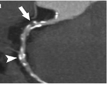
[4] Transesophageal echocardiography is an invasive procedure that involves inserting a flexible probe with an ultrasound transducer into the esophagus, providing closer access to the heart and surrounding structures.
This noninvasive and low-cost method can help diagnose and manage patients with suspected or confirmed CAD by demonstrating pathologic coronary artery flow patterns at rest and with pharmacological stress.
The technique acquires a volumetric data set and displays it in custom orientations, allowing for greater depth and understanding of heart structures compared to 2D echocardiography.
Contrast echocardiography can simultaneously assess regional myocardial function and perfusion, allowing for the non-invasive diagnosis of coronary artery disease.
[9] Faint electromagnetic signals are emitted by these hydrogen atoms when their alignment is temporarily disturbed which can be detected and used to create an image of the heart.
[10] Cardiovascular magnetic resonance (MR) technology is able to measure the size, shape, function, and tissue characteristics of the heart in a single session.
However, MR is less widely available and may be more difficult for patients to tolerate than other noninvasive modalities, requiring physician monitoring for complex cases.
[13] Recent development in deep learning and convolutional neural network techniques have made it possible to analyze and quantify some aspects of cardiac MRI automatically.
[15] The use of cardiac MRI is projected to increase through greater availability of scanners and more widespread knowledge about its clinical application.
The Wells' score for pulmonary embolism or the Diamond-Forrester chest pain criteria and Thrombolysis in Myocardial Infarction (TIMI) score can help select appropriate patients for CT.[12] Computed tomography angiography (CTA), an imaging methodology using a ring-shaped machine with an X-ray source spinning around the circular path so as to bathe the inner circle with a uniform and known X-ray density.
[16] This is achieved through the use of thin slices and high-resolution scanning, as well as the addition of electrocardiogram (ECG) gating or triggering to capture a motion-free image.
[21] PET scanners detect these gamma rays to produce images showing the location of the positrons and the metabolic processes in the body.
[21] The accuracy of the image depends on the initial speed of the emitted positron, which affects the ability of the scanner to define the position of radioactive atoms in the body.
[22] PET/MRI systems combine the capabilities of positron emission tomography (PET) and magnetic resonance imaging (MRI) to provide both functional and morphological information in various clinical applications.
[22] Cardiac MRI can produce complementary data to increase accuracy and reproducibility to PET scans, especially in systemic diseases, inflammatory processes, assessing risk of atherosclerotic plaque rupture, and stem cell tracking.
[22] Single photon emission computed tomography (SPECT), a nuclear medicine imaging methodology using gamma rays emitted by a radioactive tracer injected into the blood stream, which ultimately distributes into the heart.