Cell polarity
While the biochemical details may vary, some of the core principles such as negative and/or positive feedback between different molecules are common and essential to many known polarity systems.
Some examples of planar cell polarity include the scales of fish being oriented in the same direction and similarly the feathers of birds, the fur of mammals, and the cuticular projections (sensory hairs, etc.)
[2] Computational models have been suggested to simulate how a group of epithelial cells can form a variety of biological morphologies.
[6][7][8] The budding yeast, Saccharomyces cerevisiae, is a model system for eukaryotic biology in which many of the fundamental elements of polarity development have been elucidated.
For polarity sites to form, Cdc42 must be present and capable of cycling GTP, a process regulated by its guanine nucleotide exchange factor (GEF), Cdc24, and by its GTPase-activating proteins (GAPs).
[11] A recent study to elucidate the connection between cell cycle timing and Cdc42 accumulation in the bud site uses optogenetics to control protein localization using light.
[22] Spontaneous symmetry breaking can be explained by amplification of stochastic fluctuations of molecules due to non-linear chemical kinetics.
[24] Briefly, if a network of at least two interacting chemicals (in this case, proteins) exhibits certain types of reaction kinetics, as well as differential diffusion, stochastic concentration fluctuations can give rise to the formation of large-scale stable patterns, thus bridging from a molecular length scale to a cellular or even tissue scale.
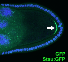
On the one hand, PAR-3, PAR-6 and aPKC (called anterior PAR proteins) occupy both the plasma membrane and cytoplasm prior to symmetry breaking.
[25] The male centrosome provides a cue, which breaks an initially homogenous membrane distribution of anterior PARs by inducing cortical flows.
[26][27] Anterior and posterior PAR proteins then maintain polarity until cytokinesis by mutually excluding each other from their respective cell membrane areas.