Cerebellar vermis
The vermis is included within the spinocerebellum and receives somatic sensory input from the head and proximal body parts via ascending spinal pathways.
[5] Inspection of the posterior fossa is a common feature of prenatal ultrasound and is used primarily to determine whether excess fluid or malformations of the cerebellum exist.
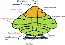
[1][7] The vermis is intimately associated with all regions of the cerebellar cortex, which can be divided into three functional parts, each having distinct connections with the brain and spinal cord.
[9] The spinocerebellum receives proprioception input from the dorsal columns of the spinal cord (including the spinocerebellar tract) and from the trigeminal nerve, as well as from visual and auditory systems.
It sends fibers to deep cerebellar nuclei that, in turn, project to both the cerebral cortex and the brain stem, thus providing modulation of descending motor systems.
Purkinje cells of the vermis project to the fastigial nucleus, controlling the axial and proximal musculature involved in the execution of limb movements.
[10] Purkinje cells in the intermediate zone of the spinocerebellum project to the interposed nuclei, which control the distal musculature components of the descending motor pathways needed for limb movement.
Upon the advent of computerized tomography (CT) and magnetic resonance imaging (MRI), the resolution of cranial structures including the mid-hindbrain regions improved dramatically.
[17] Dandy Walker malformation is characterized by enlarged posterior fossa and in which the cerebellar vermis is completely absent, or present in a rudimentary form, sometimes rotated accompanied by an elevation of the fourth ventricle.
Diagnostic features include fusion of the midbrain colliculi, hydrocephalus, absence of the corpus callosum other midline structural brain malformations.
[24] Lesions to the vermis commonly give rise to clinical depression, inappropriate emotional displays (e.g. unwarranted giggling) in addition to movement disorders.
[citation needed] Early neurophysiologists suggest that retinal and inertial signals were selected for about 450 million years ago by primitive brainstem-cerebellar circuitry because of their relationship with the environment.
[25] The origin of the cerebellum is in close association with that of the nuclei of the vestibular cranial nerve and lateral line nerves, perhaps suggesting that this part of the cerebellum originated as a means of carrying out transformations of the coordinate system from input data of the vestibular organ and the lateral line organs.
[27] In vertebrates, the cerebellar vermis develops between two bilaterally symmetrical formations located dorsal to the upper end of the medulla oblongata, or rhombencephalon.