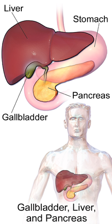
Cholecystitis
[4] Occasionally, acute cholecystitis occurs as a result of vasculitis or chemotherapy, or during recovery from major trauma or burns.
It is episodic, occurring after eating greasy or fatty foods, and leads to nausea and/or vomiting.
[13] On physical examination, an inflamed gallbladder is almost always tender to the touch and palpable (~25-50% of cases) in the midclavicular right lower rib margin.
[14] People who are old, have diabetes, chronic illness, or who are immunocompromised may have vague symptoms that may not include fever or localized tenderness.
Distension can lead to decreased blood flow to the gallbladder, causing tissue death and eventually gangrene.
[13] Once tissue has died, the gallbladder is at greatly increased risk of rupture (perforation), which can cause sharp pain.
[13] The inflammation of cholecystitis can lead to adhesions between the gallbladder and other parts of the gastrointestinal tract, most commonly the duodenum.
[13] These adhesions can lead to the formation of direct connections between the gallbladder and gastrointestinal tract, called fistulas.
Gallstones can get trapped in the gastrointestinal tract, most commonly at the connection between the small and large intestines (ileocecal valve).
[13] Gallstones are the most common cause of gallbladder inflammation but it can also occur due to blockage from a tumor or scarring of the bile duct.
[17] Risk factors for gallstones include female sex, increasing age, pregnancy, oral contraceptives, obesity, diabetes mellitus, ethnicity (Native North American), rapid weight loss.
[1] The gallbladder is initially sterile but often becomes infected by bacteria, predominantly E. coli, Klebsiella, Streptococcus, and Clostridium species.
[13] Inflammation can spread to the outer covering of the gallbladder and surrounding structures such as the diaphragm, causing referred right shoulder pain.
[13] The diagnosis of cholecystitis is suggested by the history (abdominal pain, nausea, vomiting, fever) and physical examinations in addition to laboratory and ultrasonographic testing.
[1][26][27] Ultrasound findings suggestive of acute cholecystitis include gallstones, pericholecystic fluid (fluid surrounding the gallbladder), gallbladder wall thickening (wall thickness over 3 mm),[28] dilation of the bile duct, and sonographic Murphy's sign.
[13] Given its higher sensitivity, hepatic iminodiacetic acid (HIDA) scan can be used if ultrasound is not diagnostic.
These alternative diagnoses include but are not limited to:[14] For most people with acute cholecystitis, the treatment of choice is surgical removal of the gallbladder, laparoscopic cholecystectomy.
[32] Laparoscopic cholecystectomy is performed using several small incisions located at various points across the abdomen.
[35] During the days prior to laparoscopic surgery, studies showed that outcomes were better following early removal of the gallbladder, preferably within the first week.
[36] Early laparoscopic cholecystectomy (within 7 days of visiting a doctor with symptoms) as compared to delayed treatment (more than 6 weeks) may result in shorter hospital stays and a decreased risk of requiring an emergency procedure.
[37] There is no difference in terms of negative outcomes including bile duct injury or conversion to open cholecystectomy.
[37] For early cholecystectomy, the most common reason for conversion to open surgery is inflammation that hides Calot's triangle.
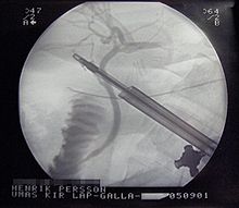
[40] In cases of severe inflammation, shock, or if the person has higher risk for general anesthesia (required for cholecystectomy), an interventional radiologist may insert a percutaneous drainage catheter into the gallbladder (percutaneous cholecystostomy tube) and treat the person with antibiotics until the acute inflammation resolves.