[1] During the later stages of cell division these chromatids separate longitudinally to become individual chromosomes.
However, if mutations occur, they will present slight differences, in which case they are heterozygous.
The pairing of chromatids should not be confused with the ploidy of an organism, which is the number of homologous versions of a chromosome.
Once sister chromatids have separated (during the anaphase of mitosis or the anaphase II of meiosis during sexual reproduction), they are again called chromosomes, each having the same genetic mass as one of the individual chromatids that made up its parent.
In chromosomal crossovers, non-sister (homologous) chromatids form chiasmata to exchange genetic material during the prophase I of meiosis (See Homologous chromosome pair).
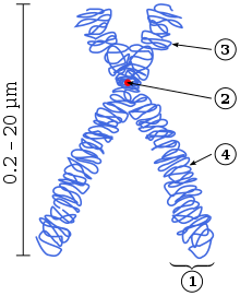
In the diagram, (1) refers to a chromatid: 1-half of two identical threadlike strands of a replicated
chromosome
. During cell division, the identical copies (called a "
sister chromatid pair
") are joined at the region called the
centromere
(2). Once the paired sister chromatids have separated from one another (in the
anaphase
of
mitosis
) each is known as a daughter chromosome. The short arm of the right chromatid (3), and the long arm of the right chromatid (4), are also marked.
Schematic
karyogram
of the human chromosomes, showing their usual state in the G
0
and G
1
phase of the cell cycle. At top center it also shows the chromosome 3 pair in
metaphase
(annotated as "Meta."), which takes place after having undergone
DNA synthesis
which occurs in the
S phase
(annotated as S) of the cell cycle. During metaphase, each chromosome is duplicated into
sister chromatids
.