Chrysosporium
[2] Chrysosporium colonies are moderately fast-growing, flat, white to tan to beige in color; they often have a powdery or granular surface texture.
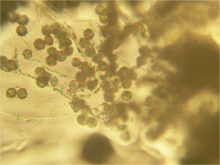
Hyaline, one-celled (ameroconidia) are produced directly on vegetative hyphae by non-specialized conidiogenous cells.
Conidia are typically pyriform to clavate with truncate bases (6 to 7 by 3.5 to 4 um) and are formed either intercalary (arthroconidia), laterally (often on pedicels), or terminally.
Species of Chrysosporium are occasionally isolated from skin and nail scrapings, especially from feet, but, because they are common soil saprotrophs, they are usually considered as contaminants.
[citation needed] Chrysosporium has been identified as an emerging infectious disease, first in Canada affecting reptiles at around 1995.