Oral candidiasis
[3] This candidal carriage state is not considered a disease, but when Candida species become pathogenic and invade host tissues, oral candidiasis can occur.
This change usually constitutes an opportunistic infection by normally harmless micro-organisms because of local (i.e., mucosal) or systemic factors altering host immunity.
Traditionally, oral candidiasis is classified using the Lehner system, originally described in the 1960s, into acute and chronic forms (see table).
[8] It is characterized by a coating or individual patches of pseudomembranous white slough that can be easily wiped away to reveal erythematous (reddened), and sometimes minimally bleeding, mucosa beneath.
[6] Frequently, antifungal therapy alone does not permanently resolve these lesions, but rather the underlying predisposing factors must be addressed, in addition to treating the candidiasis.
[4] Candida species are involved, and in some cases the lesion responds to antifungal therapy, but it is thought that other factors exist, such as oral hygiene and human herpesviruses.
[16] This is an uncommon form of chronic (more than one month in duration) candidal infection involving multiple areas in the mouth, without signs of candidiasis on other mucosal or cutaneous sites.
Unusually for candidal infections, there is an absence of predisposing factors such as immunosuppression, and it occurs in apparently healthy individuals, normally elderly males.
[13] This refers to a group of rare syndromes characterized by chronic candidal lesions on the skin, in the mouth and on other mucous membranes (i.e., a secondary oral candidiasis).
For Candida species to colonize and survive as a normal component of the oral microbiota, the organisms must be capable of adhering to the epithelial surface of the mucous membrane lining the mouth.
[13] Age also influences oral carriage, with the lowest levels occurring in newborns, increasing dramatically in infants, and then decreasing again in adults.
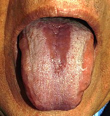
[3][23] Corticosteroid medications may contribute to the appearance of oral candidiasis,[24] as they cause suppression of immune function either systemically or on a local/mucosal level, depending on the route of administration.
[6] Candidiasis appears at the sites where the steroid has contacted the mucosa, typically the dorsum of the tongue (median rhomboid glossitis) and sometimes also on the palate.
Sometimes dentures become very worn, or they have been constructed to allow insufficient lower facial height (occlusal vertical dimension), leading to over-closure of the mouth (an appearance sometimes described as "collapse of the jaws").
This causes deepening of the skin folds at the corners of the mouth (nasolabial crease), in effect creating intertriginous areas where another form of candidiasis, angular cheilitis, can develop.
Malnutrition,[3] whether by malabsorption,[17] or poor diet, especially hematinic deficiencies (iron, vitamin B12, folic acid) can predispose to oral candidiasis,[6] by causing diminished host defense and epithelial integrity.
[9] In vitro and studies show that Candidal growth, adhesion and biofilm formation is enhanced by the presence of carbohydrates such as glucose, galactose and sucrose.
One hypothesis is that cigarette smoke contains nutritional factors for C. albicans, or that local epithelial alterations occur that facilitate colonization of candida species.
[28] Broad-spectrum antibiotics (e.g. tetracycline) eliminate the competing bacteria and disrupt the normally balanced ecology of oral microorganisms,[5][6] which can cause antibiotic-induced candidiasis.
[3] Several other factors can contribute to infection, including endocrine disorders (e.g. diabetes when poorly controlled),[30] and/or the presence of certain other mucosal lesions, especially those that cause hyperkeratosis and/or dysplasia[4] (e.g. lichen planus).
This is helpful in distinguishing pseudomembraneous candidiasis from other white lesions in the mouth that cannot be wiped away, such as lichen planus, oral hairy leukoplakia.
[31] Smears are collected by gentle scraping of the lesion with a spatula or tongue blade and the resulting debris directly applied to a glass slide.
[13] Prophylactic use of antifungals is sometimes employed in persons with HIV disease, during radiotherapy, during immunosuppressive or prolonged antibiotic therapy as the development of candidal infection in these groups may be more serious.
An alternative method of disinfection is to use a 10% solution of acetic acid (vinegar) as an overnight soak, or to microwave the dentures in 200mL water for 3 minutes at 650 watts.
It is possible for candidiasis to spread to/from the mouth, from sites such as the pharynx, esophagus, lungs, liver, anogenital region, skin or the nails.
The observation that Candida species are normally harmless commensals on the one hand, but are also occasionally capable of causing fatal invasive candidiases, has led to the description "Dr Jekyll and Mr Hyde".
[38] The role of thrush in the hospital and ventilated patients is not entirely clear, however, there is a theoretical risk of positive interaction of candida with topical bacteria.
This is due to developments in medicine, with more invasive medical procedures and surgeries, more widespread use of broad spectrum antibiotics and immunosuppression therapies.
The incidence of candidiasis caused by NCAC species is also increasing, again thought to be due to changes in medical practise (e.g., organ transplantation and use of indwelling catheters).
in "Epidemics" (a treatise that is part of the hippocratic corpus), where descriptions of what sounds like oral candidiasis are stated to occur with severe underlying disease.