Optical sectioning
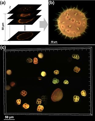
Optical sectioning is the process by which a suitably designed microscope can produce clear images of focal planes deep within a thick sample.
Oil immersion objectives typically have even larger numerical apertures so improved optical sectioning.
where λ is the wavelength, n the refractive index of the objective lens immersion media and NA the numerical aperture.
As the offset in the light sources is small the only difference in phase results from the material close to the focal plane.
This adds an extra way in which optical sectioning can be improved by making illumination specific to only the focal plane.
[5] This method effectively reduces out-of focus light and may in addition lead to a modest improvement in longitudinal resolution, compared to epi fluorescence microscopy.
The additional "concentration"-dependent effect of requiring multiple photons to simultaneously interact with a fluorophore gives stimulation only very close to the focal plane.
[6] Further improvements in optical sectioning are under active development, these principally work through methods to circumvent the diffraction limit of light.
[9] Optical sectioning can be enhanced by the use of clearing agents possessing a high refractive index (>1.4) such as Benzyl-Alcohol/Benzyl Benzoate (BABB) or Benzyl-ether[10] which render specimens transparent and therefore allow for observation of internal structures.
3D imaging using a combination of focal sectioning and tilting has been demonstrated theoretically and experimentally in order to provide exceptional 3D resolution over large fields of view.