Near-field scanning optical microscope
[5][6] As in optical microscopy, the contrast mechanism can be easily adapted to study different properties, such as refractive index, chemical structure and local stress.
His original idea, proposed in 1928, was based upon the usage of intense nearly planar light from an arc under pressure behind a thin, opaque metal film with a small orifice of about 100 nm.
He thought the moving of the pinhole or the detector when it is so close to the sample would be the most likely issue that could prevent the realization of such an instrument.
[9][10] It was Ash and Nicholls at University College London who, in 1972, first broke the Abbe's diffraction limit using microwave radiation with a wavelength of 3 cm.
[11] A decade later, a patent on an optical near-field microscope was filed by Dieter Pohl,[12] followed in 1984 by the first paper that used visible radiation for near field scanning.
[13] The near-field optical (NFO) microscope involved a sub-wavelength aperture at the apex of a metal coated sharply pointed transparent tip, and a feedback mechanism to maintain a constant distance of a few nanometers between the sample and the probe.
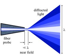
Unless the aperture of the optical component is large enough to collect all the diffracted light, the finer aspects of the image will not correspond exactly to the object.
Some types of NSOM operation utilize a campanile probe, which has a square pyramid shape with two facets coated with a metal.
[23] The merits of aperture and apertureless NSOM configurations can be merged in a hybrid probe design, which contains a metallic tip attached to the side of a tapered optical fiber.
[24] Feedback mechanisms are usually used to achieve high resolution and artifact free images since the tip must be positioned within a few nanometers of the surfaces.
In shear force feedback mode, a tuning fork is mounted alongside the tip and made to oscillate at its resonance frequency.
It is also possible to provide contrast using the change in refractive index, reflectivity, local stress and magnetic properties amongst others.
[18][19] The primary components of an NSOM setup are the light source, feedback mechanism, the scanning tip, the detector and the piezoelectric sample stage.
The light source is usually a laser focused into an optical fiber through a polarizer, a beam splitter and a coupler.
[27] The nanofocusing technique can create a nanometer-scale "white" light source at the tip apex, which can be used to illuminate a sample at near-field for spectroscopic analysis.
The interband optical transitions in individual single-walled carbon nanotubes are imaged and a spatial resolution around 6 nm has been reported.
In apertureless NSOM, also known as scattering-type SNOM or s-SNOM, many of these artifacts are eliminated or can be avoided by proper technique application.
[citation needed] An additional limitation is the predominant orientation of the polarization state of the interrogating light in the near-field of the scanning tip.