Cowpea chlorotic mottle virus
In the natural host, viral particles can be produced at 1–2 mg per gram of infected leaf tissue.
Then, under slightly acidic pH and with relatively high amounts of salts, it is possible to stimulate the self-assembly of the protein subunits, into a shell of identical size to the virus.
Due to these four species of single-stranded, positive sense RNA molecules, the CCMV genome codes for four separate genes.
[2] Lipofectamine is a reagent used in the lab to aid in transfection, allowing foreign DNA to enter the target cell.
In a study by Garmann et al. they found that the CCMV viral capsids are very robust, remaining intact even after treatment with RNase in the absence of lipofectamine.
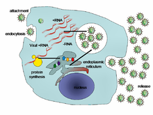
One study examined the interactions between CCMV and cowpea protoplasts and found that it was dependent on aspecific binding, mostly relying on electrostatic interactions between the plasma membrane and virus particles, specifically negatively charged vesicles and the positively charged N-terminal arm of viral coat proteins, further labelling CCMV as an endocytic virus.
The movement of CCMV requires no budding because the tubule structures enlarge the plasmodesmata enough to allow the direct passage of the viral capsid through the cell wall.
[citation needed] After virus entry, the protein capsid is degraded by the host cell, and this allows the unpackaging of the viral RNA.
[7] Upon joint infection of plant host cells with two different CCMV gene deletion mutants, functional RNA virus genomes can be regenerated by homologous recombination repair.
This process is reliant on the basicity of the CP due to its N-terminal arginine-rich motif (ARM) and the capsid exterior negative charge density.
The primarily observed symptom of CCMV is bright chlorosis, or yellow coloring, in the leaves of the plant, known as the CCMV-T strain.
This chlorosis has been observed as a less severe effect, producing a light green coloration when infecting plants with an attenuated strain, termed CCMV-M.
Results from an experiment conducted by de Assis Filho et al. indicated that this primary symptom was caused by the amino acid at position 151 of the capsid coat protein.
In this experiment, it was found that protein 1a was the only viral factor needed to induce invagination of the endoplasmic reticulum and begin the replication process.
The significance of this experiment stretches beyond the scope of the results, because S. cerevisiae is a popular model organism for viral inoculation and may open avenues for further research with CCMV.