Fluid-attenuated inversion recovery
[1] It was invented by Graeme Bydder, Joseph Hajnal, and Ian Young in the early 1990s.
By carefully choosing the inversion time (TI), the signal from any particular tissue can be nulled.
In the case of CSF suppression, one aims for T1-weighted images, which prioritize the signal of fat over that of water.
Therefore, if the long TI (inversion time) is adjusted to a zero crossing point for water (none of its signal is visible), the signal of the CSF is theoretically being "erased," from the derived image.
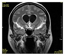
[3] The FLAIR sequence analysis has been especially useful in the evaluation and study of CNS disorders, involving:[3] * Post-contrast FLAIR images have been added to diagnosis protocol for accurate medical assessment.