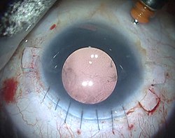
Glued intraocular lens
In ophthalmology, glued intraocular lens[1] or glued IOL is a surgical technique for implantation, with the use of biological glue, of a posterior chamber IOL (intraocular lens) in eyes with deficient or absent posterior capsules.
A quick-acting surgical fibrin sealant derived from human blood plasma,[2][3] with both hemostatic and adhesive properties, is used.
On 14 December 2007, the first glued intraocular lens (IOL) surgery was performed, at Dr. Agarwal's Eye Hospital in Chennai, India.
Subsequently, the first child on whom a glued IOL surgery was performed was a patient who had a history of injury to her right eye 3 months before, while bursting crackers.
Glued IOL surgery can be done both as a primary and as a secondary procedure in cases where the lens capsule is deficient or absent.
Fibrin glue has been used previously, in various medical specialties, as a hemostatic agent to arrest bleeding, seal tissues, and as an adjunct to wound healing.
0.5 cc of distilled water is then added to the thrombin vial, and the aprotinin is mixed with fibrinogen.
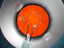
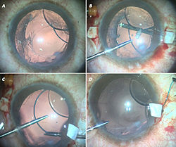
A corneal tunnel is fashioned, then a 23-gauge glued-IOL forceps is passed through the sclerotomy site, and the tip of the leading haptic of the IOL is grasped, which is then externalized and brought out onto the ocular surface (Fig 3).
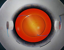
Scleral pockets are made at the edge of the flap with a 26-gauge needle just parallel to the sclerotomy site, into which the two haptics are then tucked for additional stability (Fig 4).
Advantages: According to some studies by Steven Safran, it is essential to state that the diameter of the ciliary sulcus and the corneal horizontal white-white diameter may not co-relate exactly; and it has been suggested that the surgeon can go ahead with horizontal placement of haptics rather than orienting them vertically.
Plugging Silicon Tires of Iris Hooks – This technique was developed by George Beiko and Roger Steinert, wherein the silicon tires of the iris hooks are plugged to the leading haptic, to prevent its accidental slippage.
If one of the haptics is not caught or if it is released accidentally after being grasped, the situation can be easily resolved using this technique.
This technique thus allows easy intra-ocular maneuvering of the entire haptic or IOL within a closed globe system.
Monofocal intraocular lenses, which are commonly available, give clear far or near point-of-focus, but are limited to only one focal point.
Multifocal intraocular lenses are designed to avoid the need for glasses by providing two or more points of focus.
The modified Prolene polyvinylidene fluoride haptic in these IOLs helps them in being more stiff as well as having superior structural memory.
Until recently, it was difficult to provide multifocality for patients who had complicated cataract surgeries and who lacked normal capsules.
If the endothelium is bad, the cornea retains a lot of water and becomes damaged, which is called Bullous Keratoplasty.
On 4 September 2013, Amar Agarwal, in collaboration with Harminder Dua, performed the first PDEK surgery technique and demonstrated the significance of the Pre-Descemets layer in corneal transplantation.
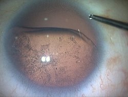
Fibrin glue takes only 20 seconds to act in the scleral bed, and it helps in adhesion and hemostasis.
