Colloidal gold
[11] Used since ancient times as a method of staining glass, colloidal gold was used in the 4th-century Lycurgus Cup, which changes color depending on the location of light source.
About half a century later, English botanist Nicholas Culpepper published a book in 1656, Treatise of Aurum Potabile,[15] solely discussing the medical uses of colloidal gold.
[17][18] In 1856, in a basement laboratory of Royal Institution, Faraday accidentally created a ruby red solution while mounting pieces of gold leaf onto microscope slides.
[22] Apart from Zsigmondy, Theodor Svedberg, who invented ultracentrifugation, and Gustav Mie, who provided the theory for scattering and absorption by spherical particles, were also interested in the synthesis and properties of colloidal gold.
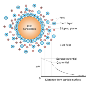
[25] Both the surface plasmon resonance frequency and scattering intensity depend on the size, shape composition and environment of the nanoparticles.
[28] In addition to solvent environment, the extinction peak can be tuned by coating the nanoparticles with non-conducting shells such as silica, biomolecules, or aluminium oxide.
[31][32][33][34][35] Colloidal gold particles can be attached to many traditional biological probes such as antibodies, lectins, superantigens, glycans, nucleic acids,[36] and receptors.
[42] The administration of hydrophobic drugs require molecular encapsulation and it is found that nanosized particles are particularly efficient in evading the reticuloendothelial system.
In cancer research, colloidal gold can be used to target tumors and provide detection using SERS (surface enhanced Raman spectroscopy) in vivo.
These gold nanoparticles are surrounded with Raman reporters, which provide light emission that is over 200 times brighter than quantum dots.
It is found that intravenously administered spherical gold nanoparticles broadened the temporal profile of reflected optical signals and enhanced the contrast between surrounding normal tissue and tumors.
[44] Gold nanoparticles have shown potential as intracellular delivery vehicles for siRNA oligonucleotides with maximal therapeutic impact.
As a consequence, for in-vivo studies, small diameter gold nanorods are being used as photothermal converters of near-infrared light due to their high absorption cross-sections.
[46] Since near-infrared light transmits readily through human skin and tissue, these nanorods can be used as ablation components for cancer, and other targets.
[47] Despite the unquestionable success of gold nanorods as photothermal agents in preclinical research, they have yet to obtain the approval for clinical use because the size is above the renal excretion threshold.
Researchers have developed simple inexpensive methods for on-site detection of hydrogen sulfide H2S present in air based on the antiaggregation of gold nanoparticles (AuNPs).
Dissolving H2S into a weak alkaline buffer solution leads to the formation of HS-, which can stabilize AuNPs and ensure they maintain their red color allowing for visual detection of toxic levels of H2S.
[54] The biocompatibility and high surface energy of Au allow it to bind to a large amount of protein without altering its activity and results in a more sensitive sensor.
[71] The possibility to use glyconanoparticles in ELISA was unexpected, but the method seems to have a high sensitivity and thus offers potential for development of specific assays for diagnostic identification of antibodies in patient sera.
[76] On the other hand, resistance to bending is found to be greatly reduced in nanoparticle monolayers that are supported at the air/water interface, possibly due to screening of ligand interactions in a wet environment.
[79] This ligand exchange can produce conjugation with a number of biomolecules from DNA to RNA to proteins to polymers (such as PEG) to increase biocompatibility and functionality.
[78] Thiolate-gold interfaces at the nanoscale have been well-studied and the thiolate ligands are observed to pull Au atoms off of the surface of the particles to form “staple” motifs that have significant Thiyl-Au(0) character.
[90] In vivo assessments can determine the general health of an organism (abnormal behavior, weight loss, average life span) as well as tissue specific toxicology (kidney, liver, blood) and inflammation and oxidative responses.
[96] However, citrate-capped gold nanoparticles sizes 8-37 nm were found to be lethally toxic for mice, causing shorter lifespans, severe sickness, loss of appetite and weight, hair discoloration, and damage to the liver, spleen, and lungs; gold nanoparticles accumulated in the spleen and liver after traveling a section of the immune system.
[102] Different sized AuNPs were found to buildup in the blood,[103][104] brain,[103] stomach,[103] pancreas,[103] kidneys,[103] liver,[103][104] and spleen.
[103][104] Biosafety and biokinetics investigations on biodegradable ultrasmall-in-nano architectures have demonstrated that gold nanoparticles are able to avoid metal accumulation in organisms through escaping by the renal pathway.
This method was discovered by Brust and Schiffrin in the early 1990s,[115] and can be used to produce gold nanoparticles in organic liquids that are normally not miscible with water (like toluene).
This approach, discovered by Perrault and Chan in 2009,[121] uses hydroquinone to reduce HAuCl4 in an aqueous solution that contains 15 nm gold nanoparticle seeds.
[125] This work pioneered the use of ultrasound to provide the energy for the processes involved and allowed the creation of gold particles with a diameter of under 10 nm.
Antibiotic functionalized metal nanoparticles have been widely studied as a mode to treat multi-drug resistant bacterial strains.