Gram stain
While Gram staining is a valuable diagnostic tool in both clinical and research settings, not all bacteria can be definitively classified by this technique.
The method is named after its inventor, the Danish scientist Hans Christian Gram (1853–1938), who developed the technique while working with Carl Friedländer in the morgue of the city hospital in Berlin in 1884.
Gram devised his technique not for the purpose of distinguishing one type of bacterium from another but to make bacteria more visible in stained sections of lung tissue.
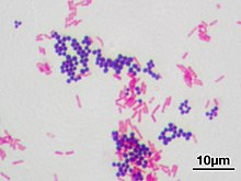
[6] Gram staining is a bacteriological laboratory technique[8] used to differentiate bacterial species into two large groups (gram-positive and gram-negative) based on the physical properties of their cell walls.
[1] Gram staining is not used to classify archaea, since these microorganisms yield widely varying responses that do not follow their phylogenetic groups.
There are four basic steps of the Gram stain: Crystal violet (CV) dissociates in aqueous solutions into CV+ and chloride (Cl−) ions.
[19] Counterstain, which is usually positively charged safranin or basic fuchsine, is applied last to give decolorized gram-negative bacteria a pink or red color.
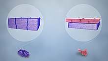
[26] Lipopolysaccharide (LPS) is the most abundant antigen on the cell surface of most gram-negative bacteria, contributing up to 80% of the outer membrane of E. coli and Salmonella.
[27] These LPS molecules, consisting of the O-antigen or O-polysaccharide, core polysaccharide, and lipid A, serve multiple functions including contributing to the cell's negative charge and protecting against certain chemicals.
LPS's role is critical in host-pathogen interactions, with the O-antigen eliciting an immune response and lipid A acting as an endotoxin.
[22] Additionally, the outer membrane acts as a selective barrier, regulated by porins, transmembrane proteins forming pores that allow specific molecules to pass.
A significant structural component linking the peptidoglycan layer and the outer membrane is Braun's lipoprotein, which provides additional stability and strength to the bacterial cell wall.
[22] Most bacterial phyla are gram-negative, including the cyanobacteria, green sulfur bacteria, and most Pseudomonadota (exceptions being some members of the Rickettsiales and the insect-endosymbionts of the Enterobacteriales).
[18][28] In cultures of Bacillus, Butyrivibrio, and Clostridium, a decrease in peptidoglycan thickness during growth coincides with an increase in the number of cells that stain gram-negative.
[33] The initial letters of gram-positive and gram-negative, which are eponymous adjectives, can be either capital G or lowercase g, depending on what style guide (if any) governs the document being written.