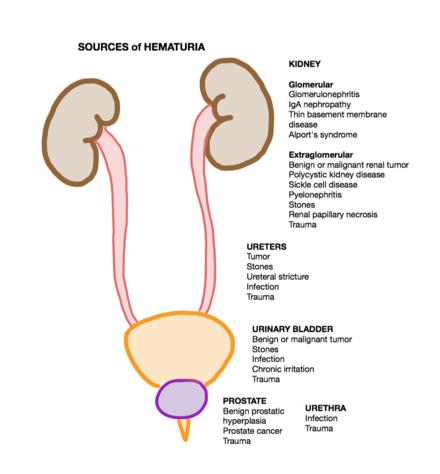
Hematuria
[3] Common causes of hematuria include urinary tract infection (UTI), kidney stones, viral illness, trauma, bladder cancer, and exercise.
A positive urine dipstick test should be confirmed with microscopy, where hematuria is defined by three or more red blood cells per high power field.
[6] When hematuria is detected, a thorough history and physical examination with appropriate further evaluation (e.g. laboratory testing) can help determine the underlying cause.
This occurs due to the red blood cells being deformed as they pass through the glomerular capillaries into the renal tubules and eventually into the urinary system.
[6] While the urine dipstick test is able to recognize heme in red blood cells, it also identifies free hemoglobin and myoglobin.
[13] It is important to obtain a detailed history from the patient (i.e. recreational, occupational, and medication exposures) as this information can be helpful in suggesting a cause of hematuria.
This medical evaluation may consist of, but is not limited too, a history and physical exam taken by healthcare personnel, laboratory studies (i.e. blood work), cystoscopy, and specialized imaging procedures (i.e. CT or MRI).
[4] In gathering information, it is important to inquire about recent trauma, urologic procedures, menses, and culture-documented urinary tract infection.
[6][3] If the results of the urinalysis and urine microscopy reveal a glomerular origin of hematuria (indicated by proteinuria or red blood cell casts), consultation with a nephrologist should be made.
[6] If the culture is positive (indicating a bladder infection), urinalysis and urine microscopy should be repeated following treatment to confirm resolution of the hematuria.
[3] After detecting and confirming hematuria with urinalysis and urine microscopy, the first step in evaluation of microhematuria is to rule out benign causes.
[15] After benign causes have resolved or been treated, a repeat urinalysis and urine microscopy is warranted to ensure cessation of hematuria.
[16] To be in the intermediate risk category, one must satisfy any of the following criteria: Has smoked 10–30 pack-years; is a female 50–59 years old or a male aged 40–59 years old; has 11–25 red blood cells per high power field; or was previously a low-risk patient with persistent microscopic hematuria and has 3–25 red blood cells per high power field.
[16] For the low risk category, the next step is to either repeat a urinalysis with urine microscopy in 6 months or perform a cystoscopy and renal ultrasound.
[18] Urosepsis is the result of a systemic inflammatory response to infection and can be identified by numerous signs and symptoms (e.g. fever, hypothermia, tachycardia, and leukocytosis).
[18] In addition to imaging tests, patients may be treated with antibiotics to relieve the infection and intravenous fluids to maintain cardiovascular and renal perfusion.
[18] Acute management of hemodynamic status, in the event intravenous fluids are unsuccessful, may include the use of vasopressor medications and the placement of a central venous line.
[19] These risks factors include age (> 40 years), male gender, previous or current smoking, chemical exposure (e.g., benzenes, hydrocarbons, aromatic amines), history of chemotherapy (alkylating agents, ifosfamide), prolonged foreign body in the bladder (such as a bladder catheter), prior pelvic radiation therapy, or greater than 25 red blood cells per high powered field on urine microscopy.


- Fill syringe with saline.
- Connect syringe to a catheter port.
- Instill 180cc of saline.
- Draw back 180cc of bladder urine.
- Dispose of medical waste.
- Repeat until all clots are removed.