Operation of computed tomography
A visual representation of the raw data obtained is called a sinogram, yet it is not sufficient for interpretation.
In terms of mathematics, the raw data acquired by the scanner consists of multiple "projections" of the object being scanned.
In conventional CT machines, an X-ray tube and detector are physically rotated behind a circular shroud (see the image above right).
An alternative, short lived design, known as electron beam tomography (EBT), used electromagnetic deflection of an electron beam within a very large conical X-ray tube and a stationary array of detectors to achieve very high temporal resolution, for imaging of rapidly moving structures, for example the coronary arteries.
In this section, how to obtain the p(s,θ) of (eq.5) by utilizing parallel beam irradiation optical system will also be explained.
Configuration and motions of parallel beam irradiation optical system, referring Fig.
Two datum coordinate systems xy and ts are imagined in order to explain the positional relations and movements of features (0)–(7) in the figure.
This datum circle (5) will be represents the orbit of the parallel beam irradiation optical system.
The μ(x,y) is absorption coefficient of the object (3) at each (x,y), p(s,θ) (7) is the collection of fluoroscopic images.
The parallel beam irradiation optical system is the key component of a CT scanner.
This datum circle (6) will be represents the orbit of the parallel beam irradiation optical system.
On the other hand, the object (1) will be scanned by CT scanner is fixed to xy coordination system.
[4] During the above-mentioned motion (that is pivoting around the object(1)) of parallel beam irradiation optical system, the collimated X-ray source (2) emits transmission beam (4) which are effectively “parallel rays” in a geometrical optical sense.
That is, transmission beam penetrates without diffraction, diffusion, or reflection although it is absorbed by the object and its attenuation is assumed to occur in accordance with the Beer-Lambert law.
More recently, manufacturers have developed iterative physical model-based maximum likelihood expectation maximization techniques.
Therefore, in this section, the explanation is advanced according to the order as follows: Consider the mathematical model where the absorption coefficient of the object at each point
Also assume the beam is absorbed by the object and its attenuation occurs in accordance with the Beer-Lambert law.
[Notes 1] around the point of origin in the plane in such a way “to keep mutual positional relationship between the light source (2) and screen (7) passing through the trajectory (5).” Rotation angle of this case is same as above-mentioned θ.
Typical implementations involve moving the patient couch through the bore of the scanner whilst the gantry rotates.
Spiral CT can achieve improved image resolution for a given radiation dose, compared to individual slice acquisition.
[8] Since its invention by Kalender in the 1980s, helical scan CT machines have steadily increased the number of rows of detectors (slices) they deploy.
A helical CT beam trajectory is characterized by its pitch, which is equal to the table feed distance along the scan range over one gantry rotation divided by the section collimation.
[10] In cone-beam computed tomography (commonly abbreviated CBCT), the X-ray beam is conical.
[11] Helical (or spiral) cone beam computed tomography is a type of three-dimensional computed tomography (CT) in which the source (usually of X-rays) describes a helical trajectory relative to the object while a two-dimensional array of detectors measures the transmitted radiation on part of a cone of rays emanating from the source.
In practical helical cone beam X-ray CT machines, the source and array of detectors are mounted on a rotating gantry while the patient is moved axially at a uniform rate.
Earlier X-ray CT scanners imaged one slice at a time by rotating source and one-dimensional array of detectors while the patient remained static.
The earliest sensors were scintillation detectors, with photomultiplier tubes excited by (typically) cesium iodide crystals.
Cesium iodide was replaced during the 1980s by ion chambers containing high-pressure xenon gas.
Initial machines would rotate the X-ray source and detectors around a stationary object.
Newer machines permitted continuous rotation with the object to be imaged slowly and smoothly slid through the X-ray ring.
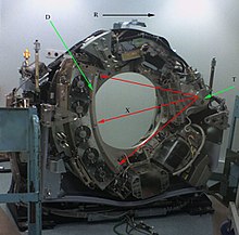
T: X-ray tube
D: X-ray detectors
X: X-ray beam
R: Gantry rotation