Scoliosis
[23] Some severe cases of scoliosis can lead to diminishing lung capacity, pressure exerted on the heart, and restricted physical activities.
[24] Longitudinal studies have revealed that the most common form of the condition, late-onset idiopathic scoliosis, causes little physical impairment other than back pain and cosmetic concerns, even when untreated, with mortality rates similar to the general population.
[25][26] Older beliefs that untreated idiopathic scoliosis necessarily progressed into severe (cardiopulmonary) disability by old age have been refuted.
[31] Adolescent idiopathic scoliosis has no clear causal agent, and is generally believed to be multifactorial; leading to "progressive functional limitations" for individuals.
[32][33][34][35][36] Research suggests that Posterior Spinal Fusion (PSF) can be used to correct the more severe deformities caused by adolescent idiopathic scoliosis.
[37][38][39][40][41] Such procedures can result in a return to physical activity in about 6 months, which is very promising, although minimal back pain is still to be expected in the most severe cases.
[citation needed] The person's gait is assessed, with an exam for signs of other abnormalities (e.g., spina bifida as evidenced by a dimple, hairy patch, lipoma, or hemangioma).
A thorough neurological examination is also performed, the skin for café au lait spots, indicative of neurofibromatosis, the feet for cavovarus deformity, abdominal reflexes and muscle tone for spasticity.
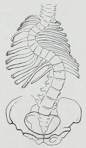
Full-length standing spine X-rays are the standard method for evaluating the severity and progression of scoliosis, and whether it is congenital or idiopathic in nature.
In growing individuals, serial radiographs are obtained at 3- to 12-month intervals to follow curve progression, and, in some instances, MRI investigation is warranted to look at the spinal cord.
[87] The traditional medical management of scoliosis is complex and is determined by the severity of the curvature and skeletal maturity, which together help predict the likelihood of progression.
[95] Significant improvement in function, vertebral angles and trunk asymmetries have been recorded following the implementation of Schroth method in terms of conservative management of scoliosis.
[89] Bracing is normally done when the person has bone growth remaining and is, in general, implemented to hold the curve and prevent it from progressing to the point where surgery is recommended.
On the opposite end of the growth spectrum, a 29° Cobb angle and a Risser sign three or four might not need to be braced because the potential for progression is reduced.
[100] Evidence supports that bracing prevents worsening of disease, but whether it changes quality of life, appearance, or back pain is unclear.
"[25][109] In an earlier University of Iowa follow-up study, 91% of people with idiopathic scoliosis displayed normal pulmonary function, and their life expectancy was found to be 2% more than that of the general population.
[26] Later (2006–) studies corroborate these findings, adding that they are "reassuring for the adult patient who has adolescent onset idiopathic scoliosis in approximately the 50–70° range.
In addition, people not having yet reached skeletal maturity have a higher likelihood of progression (i.e., if the person has not yet completed the adolescent growth spurt).
[113] Although fewer cases are present since using Cobb angle analysis for diagnosis, scoliosis remains significant, appearing in otherwise healthy children.
Despite the fact that scoliosis is a disfigurement of the spine, it has been shown to influence the pneumonic function, balance while standing and stride execution in children.
[114] Incidence of idiopathic scoliosis (IS) stops after puberty when skeletal maturity is attained, however further curvature may occur during late adulthood due to vertebral osteoporosis and weakened musculature.
In the mid-20th century, new treatments and improved screening methods have been developed to reduce the progression of scoliosis in patients and alleviate their associated pain.
[citation needed] An early treatment was the Milwaukee brace, a rigid contraption of metal rods attached to a plastic or leather girdle, designed to straighten the spine.
[citation needed] In 1962, the American orthopaedic surgeon Paul Harrington introduced a metal spinal system of instrumentation that assisted with straightening the spine, as well as holding it rigid while fusion took place.
The now obsolete Harrington rod operated on a ratchet system, attached by hooks to the spine at the top and bottom of the curvature that when cranked would distract—or straighten—the curve.
The Harrington rod obviates the need for prolonged casting, allowing patients greater mobility in the postoperative period and significantly reducing the quality of life burden of fusion surgery.
As the person aged, there would be increased wear and tear, early onset arthritis, disc degeneration, muscular stiffness, and acute pain.
[119] For instance, the human spine takes on an S-shaped curve with lumbar lordosis, which allows for better balance and support of an upright trunk.
Their fossils indicate that there may have been selected over time for a slight reduction in lumbar length to what we see today, favoring a spine that could efficiently support bipedality with a lower risk of scoliosis.
[124] Genetic testing for adolescent idiopathic scoliosis, which became available in 2009 and is still under investigation, attempts to gauge the likelihood of curve progression.