Immunogold labelling
The labeling technique can be adapted to distinguish multiple objects by using differently-sized gold particles.
[2][4] It was first applied in transmission electron microscopy (TEM) and was especially useful in highlighting proteins found in low densities, such as some cell surface antigens.
[5] As the resolution of scanning electron microscopy (SEM) increased, so too did the need for nanoparticle-sized labels such as immunogold.
In 1975, Horisberger and coworkers successfully visualised gold nanoparticles with a diameter of less than 30 nm[6] and this soon became an established SEM technique.
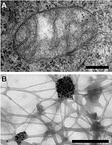
[6] The electron-dense gold particle can now be seen under an electron microscope as a black dot, indirectly labeling the molecule of interest.
[11] The silver enhancement increases the particle size, also making scanning electron microscopy possible.
An example of the application of silver-enhanced immunogold labeling (IGSS) was in the identification of the pathogen Erwinia amylovora.