Preclinical imaging
[2] These days, many manufacturers provide multi-modal systems combining the advantages of anatomical modalities such as CT and MR with the functional imaging of PET and SPECT.
[citation needed] Principle: High-frequency micro-ultrasound works through the generation of harmless sound waves from transducers into living systems.
Contrast agents can be injected into the animal to perform real-time tumor perfusion and targeted molecular imaging and quantification of biomarkers.
Principle: Photoacoustic tomography (PAT) works on the natural phenomenon of tissues to thermalelastically expand when stimulated with externally applied electromagnetic waves, such as short laser pulses.
For this reason, it can not only image structure, but also separate between different tissue types, study hemodynamic responses, and even track molecular contrast agents conjugated to specific biological molecules.
[7] To date, 3 commercially available systems are on the market, namely by VisualSonics, iThera and Endra, the last one being the only machine doing real 3D image acquisition.
For this reason, many researchers have been content to sacrifice animals at different time points and study brain tissue with traditional histological methods.
Compared to an in vivo longitudinal study, many more animals are needed to obtain significant results, and the sensitivity of the entire experiment is cast in doubt.
On the other hand, micro-fMRI is extremely expensive, and offers dismal resolution and image acquisition times when scanning the entire brain.
Micro-PAT can image the brain with high spatial resolution, detect molecular targeted contrast agents, simultaneously quantify functional parameters such as SO2 and HbT, and provide complementary information from functional and molecular imaging which would be extremely useful in tumor quantification and cell-centered therapeutic analysis.
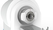
Since 2012, the use of cryogen-free magnet technology has greatly reduced infrastructure requirements and dependency on the availability of increasingly hard to obtain cryogenic coolants.
In addition, micro-MRI typically captures a snapshot of the subject in time, and thus it is unable to study blood flow and other real-time processes well.
Even with recent advances in high strength functional micro-MRI, there is still around a 10–15 second lag time to reach peak signal intensity,[10] making important information such as blood flow velocity quantification difficult to access.
[2] The X-ray is attenuated at different rates depending on the density of tissue it is passing through, and is then picked up by sensors on the opposite end of the CT scanner from the emission source.
In contrast to traditional 2D X-ray, since the emission source in a CT scanner is rotated around the animal, a series of 2D images can then be combined into 3D structures by the computer.
Although this is generally not lethal, the radiation is high enough to affect the immune system and other biological pathways, which may ultimately change experimental outcomes.
[13] Also, radiation may affect tumor size in cancer models as it mimics radiotherapy, and thus extra control groups might be needed to account for this potential confounding variable.
[14] Principle: Positron Emission Tomography (PET) images living systems by recording high-energy γ-rays emitted from within the subject.
Also, radiation may affect tumor size in cancer models as it mimics radiotherapy, and thus extra control groups might be needed to account for this potential confounding variable.
Principle: Similar to PET, single photon emission computed tomography (SPECT) also images living systems through γ-rays emitted from within the subject.
At the same time, unlike micro-PET, micro-SPECT can reach very high spatial resolution by exploring pinhole collimation principle (Beekman et al.)[16] In this approach, by placing the object (e.g. rodent) close to the aperture of the pinhole, one can reach high magnification of its projection on detector surface and effectively compensate for intrinsic resolution of the crystal.
Also, radiation may affect tumor size in cancer models as it mimics radiotherapy, and thus extra control groups might be needed to account for this potential confounding variable.
A PET-MR system provides superior soft tissue contrast and molecular imaging capability for great visualisation, quantification and translational studies.
Use of cryogen-free magnet technology also greatly reduces infrastructure requirements and dependency on the availability of increasingly hard to obtain cryogenic coolants.
In sequential PET-MR, the operator needs to allow a little time to transfer the subject between the PET and MR acquisition positions.
In sequential SPECT-MR, the operator needs to allow a little time to transfer the subject between the SPECT and MR acquisition positions.
In addition, since bioluminescence imaging does not require excitation of the reporter, but rather the catalysis reaction itself, it is indicative of the biological / molecular process and has almost no background noise.
[24][25] Since light in the infrared region has the best penetration depth, numerous fluorochromes have been specifically designed to be optimally excited in this area.
[29] Principle: Dioxaborolane chemistry enables radioactive fluoride (18F) labeling of antibodies[30] or red blood cells,[31] which allows for positron emission tomography (PET) and fluorescence imaging of cancer[32][33] and hemorrhages,[31] respectively.
Optical imaging allows for higher resolution with sub-cellular resolution of ~270 nm, or the diffraction limit of light, to allow for imaging of single cells and localizing cellular location on the cell membrane, endosomes, cytoplasm, or nuclei (see FIgure of multicolor HeLa cellls).