Lamellipodium
: lamellipodia) (from Latin lamella, related to lamina, "thin sheet", and the Greek radical pod-, "foot") is a cytoskeletal protein actin projection on the leading edge of the cell.
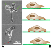
Lamellipodia are found primarily in all mobile cells, such as the keratinocytes of fish and frogs, which are involved in the quick repair of wounds.
The tip of the lamellipodium is the site where exocytosis occurs in migrating mammalian cells as part of their clathrin-mediated endocytic cycle.
[5] Another molecule that is often found in polymerizing actin with Arp2/3 is cortactin, which appears to link tyrosine kinase signalling to cytoskeletal reorganization in the lamellipodium and its associated structures.
[6] Ena/VASP proteins are found at the leading edge of lamellipodia, where they promote actin polymerization necessary for lamellipodial protrusion and chemotaxis.