Live-cell imaging
[3] The development of holotomographic microscopy has disregarded phototoxicity and other staining-derived disadvantages by implementing digital staining based on cells’ refractive index.
[4][5] Biological systems exist as a complex interplay of countless cellular components interacting across four dimensions to produce the phenomenon called life.
[6] The onerous task of capturing the true physiological identity of living tissue, therefore, requires high-resolution visualization across both space and time within the parent organism.
Due to their contiguous relationship with physiological conditions, live-cell assays are considered the standard for probing complex and dynamic cellular events.
The general acceptance of live-cell imaging has led to a rapid expansion in the number of practitioners and established a need for increased spatial and temporal resolution without compromising the health of the cell.
Phase-contrast microscopy does not have the capacity to observe specific proteins or other organic chemical compounds which form the complex machinery of a cell.
[16][17] Deep learning-assisted fluorescence microscopy methods, however, help to reduced light burden and phototoxicity and allow even repeated high resolution live imaging.
Due to the narrow focal depth of conventional microscopy, live-cell imaging is to a large extent currently limited to observing cells on a single plane.
Most implementations of quantitative phase-contrast microscopy allow creating and focusing images at different focal planes from a single exposure.
[22] Quantitative phase-contrast microscopy with rotational scanning allow 3D time-lapse images of living cells to be acquired at high resolution.
[23][24][4] Holotomography (HT) is a laser technique to measure three-dimensional refractive index (RI) tomogram of a microscopic sample such as biological cells and tissues.
Nanoscale apertures serve to calibrate the tomographic reconstruction and to characterize the imaging system by means of the coherent transfer function.
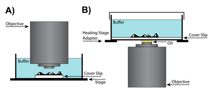
Silicone oil is an attractive medium because it has a refractive index that is close to that of living cells, allowing to produce high-resolution images while minimizing spherical aberrations.
Therefore, water-immersion lenses can help achieve a higher resolving power due to the fact that both the environment and the cells themselves will be close to the refractive index of water.
Additionally, because of the higher refractive index of water, water-immersion lenses have a high numerical aperture and can produce images superior to oil-immersion lens when resolving planes deeper than 0 μm.
[27] Today, most live imaging techniques rely on either high-illumination regimes or fluorescent labelling, both inducing phototoxicity and compromising the ability to keep cells unperturbed and alive over time.
The rise of confocal microscopy is closely correlated with accessibility of high-power lasers, which are able to achieve high intensities of light excitation.
[32] As a result, it is important to minimize the exposure of live cells to high doses of ultraviolet (UV), infrared (IR), or fluorescence exciting wavelengths of light, which can damage DNA, raise cellular temperatures, and cause photobleaching respectively.
[35] It is believed that the primary culprit in the light-induced toxicity experienced by live cells is a result of free radicals produced by the excitation of fluorescent molecules.