Physics of magnetic resonance imaging
Magnetic resonance imaging (MRI) is a medical imaging technique mostly used in radiology and nuclear medicine in order to investigate the anatomy and physiology of the body, and to detect pathologies including tumors, inflammation, neurological conditions such as stroke, disorders of muscles and joints, and abnormalities in the heart and blood vessels among other things.
Unlike CT and X-ray, MRI uses no ionizing radiation and is, therefore, a safe procedure suitable for diagnosis in children and repeated runs.
Certain atomic nuclei are able to absorb and emit radio frequency energy when placed in an external magnetic field.
In clinical and research MRI, hydrogen atoms are most often used to generate a detectable radio-frequency signal that is received by antennas close to the anatomy being examined.
Pulses of radio waves excite the nuclear spin energy transition, and magnetic field gradients localize the signal in space.
By varying the parameters of the pulse sequence, different contrasts may be generated between tissues based on the relaxation properties of the hydrogen atoms therein.
A radio frequency pulse is then applied, which can excite protons from parallel to anti-parallel alignment, only the latter are relevant to the rest of the discussion.
In response to the force bringing them back to their equilibrium orientation, the protons undergo a rotating motion (precession), much like a spun wheel under the effect of gravity.
The MRI scanner was developed from 1975 to 1977 at the University of Nottingham by Prof Raymond Andrew FRS FRSE following from his research into nuclear magnetic resonance.
[citation needed] The net longitudinal magnetization in thermodynamic equilibrium is due to a tiny excess of protons in the lower energy state.
When the radio frequency pulse is turned off, the transverse vector component produces an oscillating magnetic field which induces a small current in the receiver coil.
In an idealized nuclear magnetic resonance experiment, the FID decays approximately exponentially with a time constant T2.
A number of schemes have been devised for combining field gradients and radio frequency excitation to create an image: Although each of these schemes is occasionally used in specialist applications, the majority of MR Images today are created either by the two-dimensional Fourier transform (2DFT) technique with slice selection, or by the three-dimensional Fourier transform (3DFT) technique.
This provides high sensitivity for detection of vascular tissues (e.g., tumors) and permits assessment of brain perfusion (e.g., in stroke).
There have been concerns raised recently regarding the toxicity of gadolinium-based contrast agents and their impact on persons with impaired kidney function.
and any other phenomena that affect that amount of transverse magnetization available to induce signal in the RF probe or its phase with respect to the receiving coil' s electromagnetic field.
are conjugate variables (with respect to the Fourier transform) we can use the Nyquist theorem to show that a step in k-space determines the field of view of the image (maximum frequency that is correctly sampled) and the maximum value of k sampled determines the resolution; i.e., (These relationships apply to each axis independently.)
The second part of the pulse sequence, PE, imparts a phase shift upon the slice-selected nuclear magnetization, varying with its location in the Y direction.
During the spin echo, a frequency-encoding (FE) or readout gradient is applied, making the resonant frequency of the nuclear magnetization vary with its location in the X direction.
The negative-going lobes in GX and GZ are imposed to ensure that, at time TE (the spin echo maximum), phase only encodes spatial location in the Y direction.
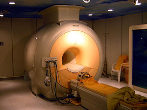
Standard foundation and comparison for other sequences Standard foundation and comparison for other sequences The major components of an MRI scanner are: the main magnet, which polarizes the sample, the shim coils for correcting inhomogeneities in the main magnetic field, the gradient system which is used to localize the MR signal and the RF system, which excites the sample and detects the resulting NMR signal.
Despite thermal insulation, sometimes including a second cryostat containing liquid nitrogen, ambient heat causes the helium to slowly boil off.
Several manufacturers now offer 'cryogenless' scanners, where instead of being immersed in liquid helium the magnet wire is cooled directly by a cryocooler.
For humans or animals the effect is particularly pronounced at air-tissue boundaries such as the sinuses (due to paramagnetic oxygen in air) making, for example, the frontal lobes of the brain difficult to image.
These are resistive coils, usually at room temperature, capable of producing field corrections distributed as several orders of spherical harmonics.
Shim currents are then adjusted to produce a large amplitude exponentially decaying FID, indicating a homogeneous B0 field.
[49] Gradient coils are used to spatially encode the positions of protons by varying the magnetic field linearly across the imaging volume.
Gradient coils are usually resistive electromagnets powered by sophisticated amplifiers which permit rapid and precise adjustments to their field strength and direction.
The RF electromagnetic radiation produced by nuclear relaxation inside the subject is true EM radiation (radio waves), and these leave the subject as RF radiation, but they are of such low power as to also not cause appreciable RF interference that can be picked up by nearby radio tuners (in addition, MRI scanners are generally situated in metal mesh lined rooms which act as Faraday cages.)
A variety of coils are available which fit closely around parts of the body such as the head, knee, wrist, breast, or internally, e.g., the rectum.