Mannose receptor
[3] The receptor recognises terminal mannose, N-acetylglucosamine and fucose residues on glycans attached to proteins [4] found on the surface of some microorganisms, playing a role in both the innate and adaptive immune systems.
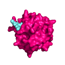
[8] The main interaction between CRD-4 and its sugar ligand is through direct ligation to the conserved Ca2+ in the sugar-binding site, in a similar way to the binding mechanism of mannan-binding lectin (MBL).
However, a quarter of the free energy of sugar-binding is associated with the hydrophobic stacking interactions formed between one face of the sugar ring and the side chain of a conserved tyrosine residue in the binding site, which is not seen in MBL.
The C-type CRDs are each separated by linker regions of 10-20 amino acids containing a number of proline residues, whose cyclic side chain is fairly rigid and favours a conformation in which the N-terminal cysteine-rich domain is extended as far away from the plasma membrane as possible.
The resistance to proteolysis shown by CRDs 4 and 5 suggests physical interactions between the two domains does occur, thereby supporting the existence of this U-shaped conformation.
The presence of sialic acid residues on N-linked glycans of the mannose receptor is important for its role in binding both sulphated and mannosylated glycoproteins.
The absence of sialic acid reduces the receptors ability to bind and internalise mannosylated glycans, but does not affect its localisation to the plasma membrane or its endocytic activity.
[6][23] The CRD regions of the mannose receptor on liver sinusoidal endothelial cells remove a number of waste material ranging from soluble macromolecules to large particulate matter.
Since the presence of mannan, an alternative ligand, inhibits LAM-dependent T cell proliferation, it is suggested that the receptor binds extracellular LAM, internalises it and then transports it to endocytic vesicles to be loaded onto CD1b.
The cleaved, soluble receptor binds to circulating antigens and directs them to effector cells in lymphoid organs via its cysteine-rich domain, thus activating the adaptive immune system.
In response to pathological events, glycoproteins including lysosomal hydrolases, tissue plasminogen activator and neutrophil myeloperoxidase are released to help fight off any invading microorganisms.
[6] High-mannose oligosaccharides present on the surface of these glycoproteins act to mark their transient nature, since they are eventually recognised by the mannose receptor and removed from the circulation.
[5] The N-terminal cysteine-rich domain of the mannose receptor plays an important role in the recognition of sulphated glycoprotein hormones and their clearance from the circulation.
Glycans on their surface are capped with sulphated N-Acetylgalactosamine (GalNAc), making them ligands for the cysteine-rich ricin homology domain of the mannose receptor.
[7] Knockout mice lacking the enzyme required to add the sulphated GalNAc capping structure show longer half-lives for lutropin, which results in increased receptor activation and oestrogen production.
Female knockout mice reach sexual maturity faster than their wild-type counterparts, have a longer oestrus cycle and produce more litters.
By manipulating the glycosylation of important bioactive proteins to a highly mannosylated state, their serum levels could be tightly regulated and they could be targeted specifically to cells expressing the mannose receptor.
[34] It has been suggested that stabilization of the Endo180-CD147 epithelial-mesenchymal transition suppressor complex and targeting of the non-complexed form of Endo180 in invasive cells could have therapeutic benefit in the prevention of cancer progression and metastasis.