Mastitis in dairy cattle
Bovine mastitis is the persistent, inflammatory reaction of the udder tissue due to physical trauma or microorganisms infections.
Mastitis, a potentially fatal mammary gland infection, is the most common disease in dairy cattle in the United States and worldwide.
Prevention and control of mastitis requires consistency in sanitizing the cow barn facilities, proper milking procedure and segregation of infected animals.
Mastitis occurs when white blood cells (leukocytes) are released into the mammary gland, usually in response to bacteria invading the teat canal or occasionally by chemical, mechanical, or thermal trauma on the udder.
Milk-secreting tissue and various ducts throughout the mammary gland are damaged due to toxins released by the bacteria resulting in reduced milk yield and quality.
[1] Microorganisms that are known to cause mastitis include: These microbes can be classified as environmental or contagious depending on mode and source of transmission.
Feeding calves on milk may introduce some mastitis causing bacteria strain in the oral cavity of the calf where it will stay dormant until it is transmitted elsewhere.
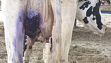
[12] Cattle affected by mastitis can be detected by examining the udder for inflammation and swelling, or by observing the consistency of the milk, which will often develop clots or change color when a cow is infected.
Coagulation of milk by rennet is sowed down due to disturbed salt balance and leucocytes reduce resazurin dye faster.
Dairy workers should wear rubber gloves while milking, and machines should be cleaned regularly to decrease the incidence of transmission.
A post milking product such as iodine-propylene glycol dip is used as a disinfectant and a barrier between the open teat and the bacteria in the air.
Mastitis can occur after milking because the teat holes close after 15 minutes if the animal sits in a dirty place with feces and urine.