Mediastinitis
Chronic sclerosing (or fibrosing) mediastinitis, while potentially serious, is caused by a long-standing inflammation of the mediastinum, leading to growth of acellular collagen and fibrous tissue within the chest and around the central vessels and airways.
Here, the fascia fuses with the pericardium and the parietal pleura, which explains the occurrence of empyema and pericardial effusion in mediastinitis.
[citation needed] Though rare in developed countries, acute mediastinitis can be caused by inhalation of bacterial spores such as Anthrax.
In the lungs, spores can spread via lymphatics to mediastinal lymph nodes, where the mature rods can release exotoxins promoting edema and tissue necrosis.
Individuals with known exposure to spores may be treated prophylactically with antibiotics (fluoroquinolones or tetracycline) to prevent disease progression.
Non-granulomatous fibrosing mediastinitis is caused by an idiopathic reaction to drugs and radiation therapy.
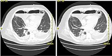
Other studies that can be used include endoscopic visualization, Chest CT scan with oral and intravenous contrast.
If discrete fluid collections or grossly infected tissue have formed (such as abscesses), they may have to be surgically drained or debrided.
[7] Treatment for chronic fibrosing mediastinitis is somewhat controversial, and may include steroids or surgical decompression of affected vessels.
The mortality of DNM ranges from 10 to 40% due to sepsis and multi-organ failure if not recognized and intervened upon early.