Multiple endocrine neoplasia
[16] The MEN1 gene consists of ten exons, spanning about 10 kb, and encodes a 610 amino acid protein named menin.
The main transcript of 2.8 kb has been described in a large variety of human tissues (pancreas, thymus, adrenal glands, thyroid, testis, leukocytes, heart, brain, lung, muscle, small intestine, liver, and kidney); an additional transcript of approximately 4 kb has been detected in pancreas and thymus, suggesting tissue-specific alternative splicing.
[17] Menin is a 610 amino acid (67Kda) nuclear protein, highly conserved from mouse (98%), rat (97%) and, more distantly, zebrafish (75%) and Drosophila (47%) (47-51).
Despite numerous studies, no genotype-phenotype correlations have been established, suggesting that unknown genetic and environmental modifiers are involved in the expression of the MEN1 phenotype.
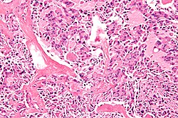
[19] Other endocrine and non-endocrine neoplasms including adrenocortical and thyroid tumors, visceral and cutaneous lipomas, meningiomas, facial angiofibromas and collagenomas, and thymic, gastric, and bronchial carcinoids also occur.
[citation needed] A recommend surveillance program for Multiple Endocrine Neoplasia Type 1 has been suggested by the International Guidelines for Diagnosis and Therapy of MEN syndromes group.
[citation needed] In 1953 Underdahl et al. reported a case series of 8 patients with a syndrome of pituitary, parathyroid, and pancreatic islet adenomas.
[citation needed] In 1961 Sipple described a combination of a pheochromocytoma, medullary thyroid carcinoma and parathyroid adenoma.
[citation needed] In 1966 Williams et al. described the combination of mucosal neuromas, pheochromocytoma and medullary thyroid carcinoma.
In 1993 mutations in the RET oncogene were shown to be the cause of MEN 2A by Lois Mulligan, working in the laboratory of Bruce Ponder in Cambridge.
[citation needed] The term "multiple endocrine neoplasia" was introduced in 1968, but descriptions of the condition date back to 1903.