Cardiac muscle
It is composed of individual cardiac muscle cells joined by intercalated discs, and encased by collagen fibers and other substances that form the extracellular matrix.
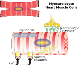
The rise in calcium causes the cell's myofilaments to slide past each other in a process called excitation-contraction coupling.
These include ischemic conditions caused by a restricted blood supply to the muscle such as angina, and myocardial infarction.
The heart wall is a three-layered structure with a thick layer of myocardium sandwiched between the inner endocardium and the outer epicardium (also known as the visceral pericardium).
On the outer aspect of the myocardium is the epicardium which forms part of the pericardial sac that surrounds, protects, and lubricates the heart.
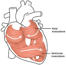
When these sheets contract in a coordinated manner they allow the ventricle to squeeze in several directions simultaneously – longitudinally (becoming shorter from apex to base), radially (becoming narrower from side to side), and with a twisting motion (similar to wringing out a damp cloth) to squeeze the maximum possible amount of blood out of the heart with each heartbeat.
[2] Contracting heart muscle uses a lot of energy, and therefore requires a constant flow of blood to provide oxygen and nutrients.
[3] They are located in the sinoatrial node (the primary pacemaker) positioned on the wall of the right atrium, near the entrance of the superior vena cava.
[6] The Purkinje fibers rapidly conduct electrical signals; coronary arteries to bring nutrients to the muscle cells, and veins and a capillary network to take away waste products.
[9] Individual cardiac muscle cells are joined at their ends by intercalated discs to form long fibers.
Each cell contains myofibrils, specialized protein contractile fibers of actin and myosin that slide past each other.
Here, a single tubule pairs with part of the sarcoplasmic reticulum, called a terminal cisterna, in a combination known as a diad.
[9] They are also involved in mechano-electric feedback,[11] as evident from cell contraction induced T-tubular content exchange (advection-assisted diffusion),[12] which was confirmed by confocal and 3D electron tomography observations.
[16][17][18][19] Under light microscopy, intercalated discs appear as thin, typically dark-staining lines dividing adjacent cardiac muscle cells.
At low magnification, this may appear as a convoluted electron dense structure overlying the location of the obscured Z-line.
In this capacity, fibroblasts can repair an injury by creating collagen while gently contracting to pull the edges of the injured area together.
When attached to a cardiomyocyte they can influence the electrical currents passing across the muscle cell's surface membrane, and in the context are referred to as being electrically coupled,[22] as originally shown in vitro in the 1960s,[23] and ultimately confirmed in native cardiac tissue with the help of optogenetic techniques.
The ECM is composed of proteins including collagen and elastin along with polysaccharides (sugar chains) known as glycosaminoglycans.
The matrix in immediate contact with the muscle cells is referred to as the basement membrane, mainly composed of type IV collagen and laminin.
The cardiac action potential subsequently triggers muscle contraction by increasing the concentration of calcium within the cytosol.
This is the direct result of a membrane which allows sodium ions to slowly enter the cell until the threshold is reached for depolarization.
[28][29] However, the mechanism by which calcium concentrations within the cytosol rise differ between skeletal and cardiac muscle.
In cardiac muscle, the action potential comprises an inward flow of both sodium and calcium ions.
[36] The complement of ion channels differs between chambers, leading to longer action potential durations and effective refractory periods in the ventricles.
[37] Diseases affecting cardiac muscle, known as cardiomyopathies, are the leading cause of death in developed countries.
[39] If these narrowings become severe enough to partially restrict blood flow, the syndrome of angina pectoris may occur.
If a coronary artery suddenly becomes very narrowed or completely blocked, interrupting or severely reducing blood flow through the vessel, a myocardial infarction or heart attack occurs.
[40] If the blockage is not relieved promptly by medication, percutaneous coronary intervention, or surgery, then a heart muscle region may become permanently scarred and damaged.
[49] Significant damage to cardiac muscle cells is referred to as myocytolysis which is considered a type of cellular necrosis defined as either coagulative or colliquative.