Muscle cell
[8] Smooth muscle cells control involuntary movements such as the peristalsis contractions in the esophagus and stomach.
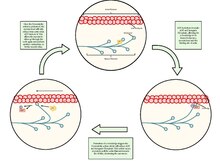
This occurs during myogenesis with the fusion of myoblasts each contributing a nucleus to the newly formed muscle cell or myotube.
The sarcoplasm also contains glycogen which provides energy to the cell during heightened exercise, and myoglobin, the red pigment that stores oxygen until needed for muscular activity.
This network is composed of groupings of two dilated end-sacs called terminal cisternae, and a single T-tubule (transverse tubule), which bores through the cell and emerge on the other side; together these three components form the triads that exist within the network of the sarcoplasmic reticulum, in which each T-tubule has two terminal cisternae on each side of it.
In the skin, smooth muscle cells such as those of the arrector pili cause hair to stand erect in response to cold temperature or fear.
Although smooth muscle cells lack sarcomeres and myofibrils they do contain large amounts of the contractile proteins actin and myosin.
The action potential uses transverse tubules to get from the surface to the interior of the myocyte, which is continuous within the cell membrane.
With a singular neuromuscular junction, each muscle fiber receives input from just one somatic efferent neuron.
Very quickly Ca2+ is actively transported back into the sarcoplasmic reticulum, which blocks the interaction between the thin and thick filament.
Specialized cardiomyocytes in the sinoatrial node generate electrical impulses that control the heart rate.
Sinoatrial node activity is modulated, in turn, by nerve fibers of both the sympathetic and parasympathetic nervous systems.
In this case, Schmid & Seipel argue that the last common ancestor of Bilateria, Ctenophora and Cnidaria, was a triploblast (an organism having three germ layers), and that diploblasty, meaning an organism with two germ layers, evolved secondarily, because of their observation of the lack of mesoderm or muscle found in most cnidarians and ctenophores.
They use an example of the contractile elements present in the Porifera, or sponges, that do truly lack this striated muscle containing this protein.
Steinmetz, Kraus, et al. showed that the traditional morphological and regulatory markers such as actin, the ability to couple myosin side chains phosphorylation to higher concentrations of the positive concentrations of calcium, and other MyHC elements are present in all metazoans not just the organisms that have been shown to have muscle cells.
This separation of the duplicated set of genes is shown through the localization of the striated much to the contractile vacuole in sponges, while the non-muscle much was more diffusely expressed during developmental cell shape and change.
[31] Steinmetz, Kraus, et al. (2012)[31] further argue for multiple origins of striated muscle in the metazoans by explaining that a key set of genes used to form the troponin complex for muscle regulation and formation in bilaterians is missing from the cnidarians and ctenophores, and 47 structural and regulatory proteins observed, Steinmetz, Kraus, et al. were not able to find even on unique striated muscle cell protein that was expressed in both cnidarians and bilaterians.
Furthermore, the Z-disc seemed to have evolved differently even within bilaterians and there is a great deal of diversity of proteins developed even between this clade, showing a large degree of radiation for muscle cells.
Through this divergence of the Z-disc, Steinmetz, Kraus, et al. argue that there are only four common protein components that were present in all bilaterians muscle ancestors and that of these for necessary Z-disc components only an actin protein that they have already argued is an uninformative marker through its pleisiomorphic state is present in cnidarians.
Through further molecular marker testing, Steinmetz et al. observe that non-bilaterians lack many regulatory and structural components necessary for bilaterians muscle formation and do not find any unique set of proteins to both bilaterians and cnidarians and ctenophores that are not present in earlier, more primitive animals such as the sponges and amoebozoans.
[32] In their paper, Andrikou & Arnone (2015)[32] argue that to truly understand the evolution of muscle cells the function of transcriptional regulators must be understood in the context of other external and internal interactions.
Through their analysis, Andrikou & Arnone found that there were conserved orthologues of the gene regulatory network in both invertebrate bilaterians and cnidarians.
They argue that having this common, general regulatory circuit allowed for a high degree of divergence from a single well-functioning network.
Andrikou & Arnone found that the orthologues of genes found in vertebrates had been changed through different types of structural mutations in the invertebrate deuterostomes and protostomes, and they argue that these structural changes in the genes allowed for a large divergence of muscle function and muscle formation in these species.
However, Andrikou & Arnone explain that the basic muscle patterning structure must also be considered in combination with the cis regulatory elements present at different times during development.
In contrast with the high level of gene family apparatuses structure, Andrikou and Arnone found that the cis-regulatory elements were not well conserved both in time and place in the network which could show a large degree of divergence in the formation of muscle cells.
[32] Evolutionarily, specialized forms of skeletal and cardiac muscles predated the divergence of the vertebrate / arthropod evolutionary line.
[34] To further complicate this classification scheme, the mitochondrial content, and other morphological properties within a muscle fiber, can change in a tsetse fly with exercise and age.