Nanoscale secondary ion mass spectrometry
[3] The original design of the NanoSIMS instrument was conceived by Georges Slodzian at the University of Paris Sud in France and at the Office National d'Etudes et de Recherches Aérospatiales.
The main drawback of this set up is that the primary and secondary ion beams must be of opposite polarity which can limit which elements can be detected simultaneously.
[5][7] The relatively large number of masses helps eliminate measurement errors as possible changes in instrumental or sample conditions that may occur in between runs are avoided.
Unlike other imaging techniques, where 13C14N and 12C15N cannot be independently measured due to nearly identical masses, NanoSIMS can safely distinguish the differences between these molecules allowing isotopic spiking experiments to be conducted.
The Lorentz force states that a particle will experience a force when it maintains a charge q and travels through an electric field E and magnetic field B with a velocity v. The secondary ions that leave the surface of the sample typically have a kinetic energy of a few electron volts (eV), although a rather small portion have been found to have energy of a few keV.
The probability that any ion will be successfully transferred from mass spectrometer to detector is T. The product of Yi and T determines the amount of isotopes that will be ionized, as well as detected, so it is considered the useful yield.
As the NanoSIMS operates under ultra high vacuum, the sample must be vacuum compatible (i.e., volatile free), flat, which reduces varying ionization trajectories, and conductive, which can be accomplished by sputter coating with Au, Pt, or C. Biological samples, such as cells or tissue, can be prepared with chemical fixation or cryo-fixation and embedded in a resin before sectioning into thin slices (100 nm - 1μm), and placed on silicon wafers or slides for analysis.
[4] NanoSIMS can capture the spatial variability of isotopic and elemental measurements of sub-micron areas, grains or inclusions from geological, materials science and biological samples.
[12] This instrument can characterise nanostructured materials with complex composition that are increasingly important candidates for energy generation and storage.
NanoSIMS has also proved useful in studying cosmochemical issues, where samples of single, micro- or sub-micrometer-sized grains from meteorites as well as microtome sections prepared by the focused ion beam (FIB) technique can be analyzed.
[13] Initially developed for geochemical and related research, NanoSIMS is now utilized by a wide variety of fields, including biology and microbiology.
This study was the first time that carbon and nitrogen isotope ratios were directly measured at a sub-cellular scale in a biological sample.
[16] The development of NanoSIMS for organo-metallic drugs paved the way for exploring the distribution of biologically active molecules at the subcellular level.
Legin et al.[17] combined NanoSIMS with fluorescence confocal laser scanning microscopy to characterize the subcellular distribution of 15N isotopically labeled Pt-bearing cisplatin in human colon cancer cells.
He et al.[19] visualized the distribution of therapeutic antisense oligonucleotides labelled with bromine (Br-ASO) in some varieties of cultured cells and importantly mouse tissues (heart, kidney, and Liver) using NanoSIMS data combined with back scattered electron microscopy.
The NanoSIMS instrument can detect the gold particles, providing the location of the labelled proteins at a high scale resolution.
Previous quantitative analysis techniques at a lower resolution than NanoSIMS of stable isotopically labeled molecules was limited to analyzed bulk material, which did not allow for insights about the contributions of individual cells or subcellular compartments to be made.
[32] This type of analysis was first used in 2001 in conjunction with FISH to examine syntrophic relationships between anaerobic methane-oxidizing archaea and sulfate reducing bacteria.
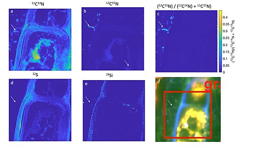
[8] NanoSIMS was first used in this field of paleobiology in 2005 by Robert et al.[34] In this study, microfossils were found to contain carbon, nitrogen, and sulfur elements arranged as ‘globules’ that were reminiscent of cell walls.