Nasal reconstruction using a paramedian forehead flap
[1] In this operation a reconstructive surgeon uses skin from the forehead above the eyebrow and pivots it vertically to replace missing nasal tissue.
Throughout history the technique has been modified and adjusted by many different surgeons and it has evolved to become a popular way of repairing nasal defects.
[4] If the defect is small and superficial it can be resurfaced with a skin graft or it can heal by secondary intention.
The forehead flap is known as the best donor site for repairing nasal defects because of its size, superior vascularity, skin color, texture and thickness.
Vascularisation of the scalp and forehead is supplied by the supraorbital, supratrochlear, superficial temporal, postauricular and occipital vessels.
[1][4][6] All these vessels are lined vertically and permit safe and effective transfer of the forehead flap on multiple individual vascular pedicles.
The perfusion of the paramedian forehead flap comes from three sources: randomly, through the frontalis muscle and through the supratrochlear artery.
[4] However, the vertical paramedian forehead flap based on the ipsilateral or contralateral supratrochlear vessels has become standard, because it has a low turning point, making it easy to reach the defect without using hair-bearing scalp.
These regions (forehead, cheeks, eyelids, lips, nose and chin) are defined by skin quality, border outline, and three-dimensional contour.
If enlarging the defect will make the aesthetic result better, normal tissue within the subunit can be safely removed.
[4] Concha, septum or rib cartilage grafts should be used for creating enough support and a good shape.
Any possible resultant defect is high in the forehead and left to heal by secondary intention.
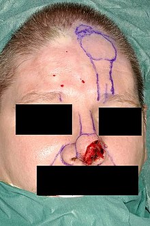
Important landmarks are the hairline, frown lines, location of the supratrochlear vessels, outline of the defect, nasal and lip subunits.
The template resembling the defect is placed just under the hairline and the vascular pedicle is drawn downwards into the medial eyebrow.
The scar is eventually sculpted between the nasal subregions to create a satisfying aesthetic result.
[1] The flap consists of skin, subcutaneous tissue, fat and frontalis muscle and is not thinned.
Three to four weeks later, when the full thickness forehead flap is well healed at the recipient site, the second stage begins.
[7] The results of nasal reconstruction using the paramedian forehead flap are quite good, although some patients report functional difficulties.
[5] Ideally, standardized semistructered interviews are used to assess aesthetic outcome after nasal reconstruction.
Studies using these interviews showed that generally patients are very satisfied with the result although they reported aggravating of their nasal appearance compared to before surgery.
[5] Remarkably, patients scored subjective aesthetic outcome significantly higher compared to a professional panel.
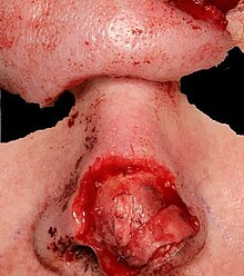
Early discovery of infection is very important so complete debridement can take place before underlying cartilage is exposed.
A second flap can be taken from the contra lateral side in most instances [1] The defect created at the donor site is usually positioned at the central/lateral forehead.
The defect can be closed by pulling the different sides of the wound together in vertical and horizontal direction.
[1] If there is a resulting defect after closure it is situated high in the forehead and it closes by secondary healing.
That is why there is a significant risk of superior eyebrow malposition, especially if a horizontal or oblique oriented forehead flap is used.