Autopsy
An autopsy (also referred to as post-mortem examination, obduction, necropsy,[Note 1] or autopsia cadaverum) is a surgical procedure that consists of a thorough examination of a corpse by dissection to determine the cause, mode, and manner of death; or the exam may be performed to evaluate any disease or injury that may be present for research or educational purposes.
Critics, including pathologist and former JAMA editor George D. Lundberg, have charged that the reduction in autopsies is negatively affecting the care delivered in hospitals, because when mistakes result in death, they are often not investigated and lessons, therefore, remain unlearned.
[7] Organizations such as ZAKA in Israel and Misaskim in the United States generally guide families on how to ensure that an unnecessary autopsy is not made.
Autopsies are used in clinical medicine to identify a medical error or a previously unnoticed condition that may endanger the living, such as infectious diseases or exposure to hazardous materials.
[10] However, this rate has decreased over time and the study projects that in a contemporary US institution, 8.4% to 24.4% of autopsies will detect major diagnostic errors.
A large meta-analysis suggested that approximately one-third of death certificates are incorrect and that half of the autopsies performed produced findings that were not suspected before the person died.
[12] Focusing on intubated patients, one study found "abdominal pathologic conditions – abscesses, bowel perforations, or infarction – were as frequent as pulmonary emboli as a cause of class I errors.
They are performed to gain more insight into pathological processes and determine what factors contributed to a patient's death.
Postmortem examinations require the skill to connect anatomic and clinical pathology together since they involve organ systems and interruptions from ante-mortem and post-mortem.
These academic autopsies allow for students to practice and develop skills in pathology and become meticulous in later case examinations.
[21] This method is helpful in determining the questions pertaining to an autopsy without putting the examiner at risk of biohazardous materials that can be in an individual's body.
[22] The rate of consented (hospital) autopsy in the UK and worldwide has declined rapidly over the past 50 years.
Toxicology, biochemical tests or genetic testing/molecular autopsy often supplement these and frequently assist the pathologist in assigning the cause or causes of death.
At many institutions, the person responsible for handling, cleaning, and moving the body is called a diener, the German word for servant.
In the UK this role is performed by an Anatomical Pathology Technician (APT), who will also assist the pathologist in eviscerating the body and reconstruction after the autopsy.
Ultraviolet light may also be used to search body surfaces for any evidence not easily visible to the naked eye.
In all of the above cases, the incision then extends all the way down to the pubic bone (making a deviation to either side of the navel) and avoiding, where possible, transecting any scars that may be present.
Bleeding from the cuts is minimal, or non-existent because the pull of gravity is producing the only blood pressure at this point, related directly to the complete lack of cardiac functionality.
When the autopsy is completed, the incision can be neatly sewn up and is not noticed when the head is resting on a pillow in an open casket funeral.
The skull is then cut with a circular (or semicircular) bladed reciprocating saw to create a "cap" that can be pulled off, exposing the brain.
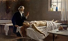
An important component of the autopsy is the reconstitution of the body such that it can be viewed, if desired, by relatives of the deceased following the procedure.
[1][32] Autopsies that opened the body to determine the cause of death were attested at least in the early third millennium BCE, although they were opposed in many ancient societies where it was believed that the outward disfigurement of dead persons prevented them from entering the afterlife[33] (as with the Egyptians, who removed the organs through tiny slits in the body).
[1] Notable Greek autopsists were Erasistratus and Herophilus of Chalcedon, who lived in 3rd century BCE Alexandria, but in general, autopsies were rare in ancient Greece.
[1] The greatest ancient anatomist was Galen (CE 129– c. 216), whose findings would not be challenged until the Renaissance over a thousand years later.
[36] The dissection of human remains for medical or scientific reasons continued to be practiced irregularly after the Romans, for instance by the Arab physicians Avenzoar and Ibn al-Nafis.
In Europe they were done with enough regularity to become skilled, as early as 1200, and successful efforts to preserve the body, by filling the veins with wax and metals.
[38] In the mid-1800s, Carl von Rokitansky and colleagues at the Second Vienna Medical School began to undertake dissections as a means to improve diagnostic medicine.
[39] During the turn of the 20th century, the Scotland Yard created the Office of the Forensic Pathologist, a medical examiner trained in medicine, charged with investigating the cause of all unnatural deaths, including accidents, homicides, suicides, etc.
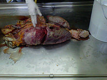
For many species that exhibit few external symptoms (sheep), or that are not suited to detailed clinical examination (poultry, cage birds, zoo animals), it is a common method used by veterinary physicians to come to a diagnosis.
The entire body is examined at the gross visual level, and samples are collected for additional analyses.