Nephron
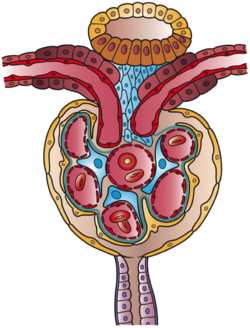
[1]: 22 Blood is filtered as it passes through three layers: the endothelial cells of the capillary wall, its basement membrane, and between the podocyte foot processes of the lining of the capsule.
The filtering structure (glomerular filtration barrier) has three layers composed of endothelial cells, a basement membrane, and podocyte foot processes.
Filtration or ultrafiltration occurs in the glomerulus and is largely passive: it is dependent on the intracapillary blood pressure.
A countercurrent system in the renal medulla provides the mechanism for generating a hypertonic interstitium, which allows the recovery of solute-free water from within the nephron and returning it to the venous vasculature when appropriate.
A nephron is made of two parts: The renal corpuscle is the site of the filtration of blood plasma.
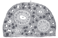
The glomerulus is the network, known as a tuft, of filtering capillaries located at the vascular pole of the renal corpuscle in Bowman's capsule.
The diameter of the efferent arteriole is smaller than that of the afferent, and this difference increases the hydrostatic pressure in the glomerulus.
The renal tubule is a continuous and long pipe-like structure containing the tubular fluid filtered through the glomerulus.
[7] The components of the renal tubule are: The epithelial cells that form these nephron segments can be distinguished by the shapes of their actin cytoskeleton visualized by confocal microscopy of fluorescent phalloidin.
These long loops of Henle and their associated vasa recta create a hyperosmolar gradient that allows for the generation of concentrated urine.
[11] Juxtamedullary nephrons are found only in birds and mammals, and have a specific location: medullary refers to the renal medulla, while juxta (Latin: near) refers to the relative position of the renal corpuscle of this nephron - near the medulla, but still in the cortex.
[12] Fluid in the filtrate entering the proximal convoluted tubule is reabsorbed into the peritubular capillaries, including 80% of glucose, more than half of the filtered salt, water and all filtered organic solutes (primarily glucose and amino acids).
The hypertonicity of the medulla (and therefore concentration of urine) is determined in part by the size of the loops of Henle.
The ascending limb actively pumps sodium out of the filtrate, generating the hypertonic interstitium that drives countercurrent exchange.
Cells lining the tubule have numerous mitochondria to produce enough energy (ATP) for active transport to take place.
Much of the ion transport taking place in the distal convoluted tubule is regulated by the endocrine system.
In the presence of parathyroid hormone, the distal convoluted tubule reabsorbs more calcium and secretes more phosphate.
The collecting duct system begins in the renal cortex and extends deep into the medulla.
ADH affects the function of aquaporins, resulting in the reabsorption of water molecules as it passes through the collecting duct.
Aquaporins are membrane proteins that selectively conduct water molecules while preventing the passage of ions and other solutes.
[5]: 406 Lower portions of the collecting organ are also permeable to urea, allowing some of it to enter the medulla, thus maintaining its high concentration (which is very important for the nephron).
It produces and secretes into the circulation the enzyme renin (angiotensinogenase), which cleaves angiotensinogen and results in the ten amino acid substance angiotensin-1 (A-1).

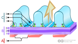
B. Glomerular basement membrane : 1. lamina rara interna 2. lamina densa 3. lamina rara externa
C. Podocytes: 1. enzymatic and structural proteins 2. filtration slit 3. diaphragm