Oogonium
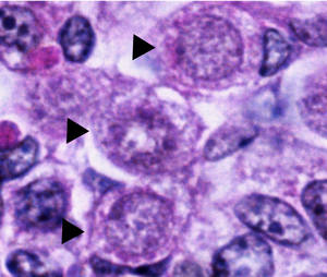
Normal oogonia in human ovaries are spherical or ovoid in shape and are found amongst neighboring somatic cells and oocytes at different phases of development.
Oogonial nuclei contain randomly dispersed fibrillar and granular material whereas the somatic cells have a more condensed nucleus that creates a darker outline under the microscope.
The chromosomal material in the nucleus of mitotically dividing oogonia shows as a dense mass surrounded by vesicles or double membranes.
Degenerating oogonia are usually found partially or wholly engulfed in neighboring somatic cells, identifying phagocytosis as the mode of elimination.
[1] In the blastocyst of the mammalian embryo, primordial germ cells arise from proximal epiblasts under the influence of extra-embryonic signals.
These germ cells then travel, via amoeboid movement, to the genital ridge and eventually into the undifferentiated gonads of the fetus.
[3][4] During the 6th to 8th week of female (XX) embryonic development, the primordial germ cells grow and begin to differentiate into oogonia.
Research has shown that ovaries lacking Rspo1 or Wnt4 will exhibit sex reversal of the gonads, the formation of ovotestes and the differentiation of somatic sertoli cells, which aid in the development of sperm.
One major factor that contributes to the up-regulation of Stra8, is the initiation of the β-Catenin signaling pathway via RSPO1, which is also responsible for ovary differentiation.
[4] It is theorized that oogonia either degenerate or differentiate into primary oocytes which enter oogenesis and are halted in prophase I of the first meiosis post partum.
[2] Recent research, however, has identified that renewable oogonia may be present in the lining of the female ovaries of humans, primates and mice.
These mitotically active germ cells found in mammalian adults were identified by tracking several markers that were common in oocytes.
[2] The discovery of these active germ cells and oogonia in the adult female could be very useful in the advancement of fertility research and treatment of infertility.
Multiple approaches to verify the existence of oogonial stem cells have yielded negative results, and no research group in United States has been able to reproduce initial findings.
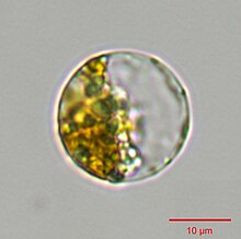
[12][13] In Oomycota and some other organisms, the female oogonia, and the male equivalent antheridia, are a result of sexual sporulation, i.e. the development of structures within which meiosis will occur.