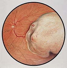
Uveal melanoma
Other risk factors, such as blue light exposure and arc welding, have been put forward, but are still debated in the field.
Approximately 50 percent of patients will develop metastases within 15 years after treatment of the primary tumor, and the liver will be involved 90% of the time.
[23] Metastasis can occur more than 10 years after treatment of the primary tumor, and patients should not be considered cured even after a 10-year interval of monitoring.
The disease-free interval, the performance status, the liver substitution by metastases and the serum level of lactic dehydrogenase are the most important prognostic factors for metastatic uveal melanoma.
Primary treatment can involve removal of the affected eye (enucleation); however, this is now reserved for cases of extreme tumor burden or other secondary problems.
Advances in radiation therapies have significantly decreased the number of patients treated by enucleation in developed countries.
The risk of metastasis after plaque radiotherapy is the same as that of enucleation, suggesting that micrometastatic spread occurs prior to treatment of the primary tumor.
Proton therapy delivers powerful doses of radiation to the tumor while sparing surrounding healthy eye tissue.
On January 25, 2022, the FDA approved tebentafusp (Kimmtrak) for adult HLA-A*2-positive patients with metastatic uveal melanoma.
[28] Ocular melanoma expert Professor Sarah Coupland, in 2018, suggested cautious optimism as new types of targeted therapeutics are tested and approved.
[30][31] Likewise several histological and cytological factors are associated with higher risk of metastasis, including presence and extent of cells with epithelioid morphology, presence of looping extracellular matrix patterns, increased infiltration of immune cells,[21] and staining with several immunohistochemical markers.
[34] In rare instances, monosomy 3 tumors may duplicate the BAP1-mutant copy of the chromosome to return to a disomic state referred to as isodisomy.
[36] Monosomy 3, along with other chromosomal gains, losses, amplifications, and LOH, can be detected in fresh or paraffin-embedded samples by virtual karyotyping.
[37][38] Gene expression profiling outperforms all of the above-mentioned factors at predicting metastatic spread of the primary tumor, including monosomy 3.
The scientific community is currently working to develop guidelines, but until then, each patient must take into consideration their individual clinical situation and discuss appropriate surveillance with their doctors.
[42] Some ophthalmologists have also found promise with the use of intravitreal avastin injections in patients with radiation-induced retinopathy, a side effect of plaque brachytherapy treatment, as well as imaging surveillance with SD-OCT. Uveal melanomas are the most common primary intraocular tumor in adults.