Retinoblastoma
Retinoblastoma (Rb) is a rare form of cancer that rapidly develops from the immature cells of a retina,[2] the light-detecting tissue of the eye.
[3] It is the most common primary malignant intraocular cancer in children, and 80% of retinoblastoma cases are first detected in those under 3 years old.
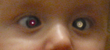
During a red reflex test, light from an ophthalmoscope goes through transparent parts of the eye and reflects off the ocular fundus.
[9] The most common and obvious sign of retinoblastoma is an abnormal appearance of the retina as viewed through the pupil, the medical term for which is leukocoria, also known as amaurotic cat's eye reflex.
[5] Other signs and symptoms include deterioration of vision, a red and irritated eye with glaucoma, and faltering growth or delayed development.
Retinoblastoma presents with advanced disease in developing countries and eye enlargement is a common finding.
[15] The defective RB1 gene can be inherited from either parent; in some children, however, the mutation occurs in the early stages of fetal development.
[17] A 2014 meta-analysis showed that 5-year survival of trilateral retinoblastoma increased from 6% before 1995 to 57% by 2014, attributed to early detection and improved chemotherapy.
[23] Somatic amplification of the MYCN oncogene is responsible for some cases of nonhereditary, early-onset, aggressive, unilateral retinoblastoma.
MYCN can act as a transcription factor and promotes proliferation by regulating the expression of cell cycle genes.
[24][25] Although MYCN amplification accounted for only 1.4% of retinoblastoma cases, researchers identified it in 18% of infants diagnosed at less than 6 months of age.
[citation needed] Gross and microscopic appearances of retinoblastoma are identical in both hereditary and sporadic types.
CT scan is generally avoided because radiation can stimulate the formation of more eye tumours in those with RB1 genetic mutation.
[38] The system was originally used to decide the best treatment result by using external beam radiotherapy, as well as, the likeliness of salvaging the globe of the eye.
[38] According to Reese and Ellsworth, there were different groups that had various features in order to classify the globe salvage as very favorable to the category of very unfavorable.
If the tumor was around ten in disc diameter and involved roughly 50% of the retina, it was considered unfavorable to salvage the globe which could result in enucleation.
When a patient is very high risk, the tumor presents itself with multiple features and is going to have to be treated with conservative treatment modalities or enucleation.
The exact course of treatment depends on the individual case and is decided by the ophthalmologist in discussion with the paediatric oncologist.
[53] A higher incidence is noted in developing countries, which has been attributed to lower socioeconomic status and the presence of human papilloma virus sequences in the retinoblastoma tissue.