Choroidal nevus
Dr. Gass, one of the leading specialists on eye diseases, speculates that a choroidal nevus grows from small cells resting as hyperplastic lesions, and exhibits growth primarily.
Choroidal nevus is usually diagnosed through an ophthalmic eye examination, or more specialized technologies such as photographic imaging, ophthalmoscopy, ultrasonography and ocular coherence tomography (OCT).
[4] In terms of ethnicity, a cohort study done in the United States reported that the prevalence of choroidal nevus was found more in whites (4.1%) than in Chinese (0.4%), blacks (0.7%) and Hispanics (1.2%).
Another study on the prevalence of choroidal nevus among the female population investigated the role of obesity and reproductive factors in the development of the disease.
When a choroidal nevus becomes severe, it can cause leakage of fluid and abnormal development of vascular tissue[9] (neovascularization[10]).
This leads to retinal detachment in that part of the eye, which is observed as some loss of vision or flashing lights.
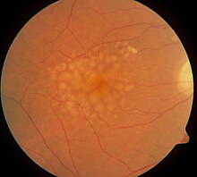
[9] Additionally, if the nevus is present for an extended period of time (years), and hinders the removal of retinal waste products, this can result in the development of yellowish white specks and spots on the surface of the nevi,[9] called drusen.
[12][11] A few indicators of a halo nevus include an absence of subretinal fluid and orange pigment, thickness level less than 2 mm, as well as the tumor margin being remote from the optic disk.
[13] Due to its large basal diameter and thickness, it can be easily mistaken and diagnosed as choroidal melanoma.
[14] Drusen are composed of lipids and can actually be an indicator that a tumour is a benign nevus as opposed to a cancerous melanoma.
These include light iris color, generally lower levels of melanin (light and untanned skin tones), exposure to arc welding due to intermittent ultraviolet exposure,[22][23] as well as diseases such as ocular melanocytosis and dysplastic nevus syndrome.
[24] In contrast, for small melanomas, the speed of growth is much faster,[13][24] making it easily detectable in a short period of time.
[24] Slow growth and enlargement of choroidal nevi are found to be more common in younger patients, before becoming stable in mid or late adulthood.
With a thickness of approximately 2mm and a color between brown to slate gray, the edge of the nevus blends into the retina.
[17] A B-scan ultrasound provides the practitioner with an approximate size of the tumor, in addition to vertical and horizontal measurements,[14] while an A-scan determines the amount of internal reflectivity.
This can be achieved through machine learning, whereby a large dataset of imaging photographs of all sizes, shapes and location of nevi are used in training.