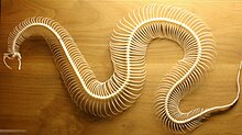
Snake skeleton
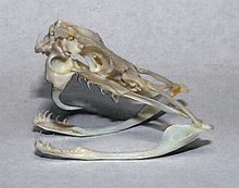
The quadrate and maxillary and palatopterygoid arches are more or less movable to allow for the distension required by the passage of prey, often much exceeding the size of the mouth.
For the same reason, the rami of the lower jaw, which consist of dentary, splenial, angular, and articular elements, with the addition of a coronoid in the boas and a few other small families, are connected at the symphysis by a very extensible elastic ligament.
A large hole may be present between the frontal bones and the basisphenoid (Psammophis, Coelopeltis); the maxillary may be much abbreviated and movable vertically, as in the Viperidae; the pterygoids may taper and converge posteriorly, without any connection with the quadrate, as in the Amblycephalidae; the supratemporal may be much reduced, and wedged in between the adjacent bones of the cranium; the quadrate may be short or extremely large; the prefrontals may join in a median suture in front of the frontals; the dentary may be freely movable, and detached from the articular posteriorly.
The deviation from the normal type is much greater still when we consider the degraded wormlike members of the families Typhlopidae and Glauconiidae, in which the skull is very compact and the maxillary much reduced.
In the former this bone is loosely attached to the lower aspect of the cranium; in the latter, it borders the mouth and is suturally joined to the premaxillary and the prefrontal.
In most snakes, teeth are located on the dentary of the lower jaw, the maxilla, the palatine bone and the lateral pterygoid plate.
Several snake lineages have evolved venom which is typically delivered by specialized teeth called fangs located on the maxilla.
Proteroglyphous snakes (forward grooved) have shortened maxillae bearing few teeth except for a substantially enlarged fang pointing downwards and completely folded around the venom channel, forming a hollow needle.
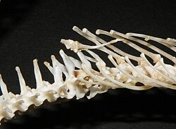
The centra have the usual ball and socket joint, with the nearly hemispherical or transversely elliptic condyle at the back (procoelous vertebrae), while the neural arch is provided with additional articular surfaces in the form of pre- and post-zygapophyses, broad, flattened, and overlapping, and of a pair of anterior wedge-shaped processes called zygosphene, fitting into a pair of corresponding concavities, zygantrum, just below the base of the neural spine.
The precaudal vertebrae have a more or less high neural spine which, as a rare exception (Xenopholis), may be expanded and plate-like above, and short or moderately long transverse processes to which the ribs are attached by a single facet.
The centra of the anterior vertebrae emit more or less developed descending processes, or haemapophyses, which are sometimes continued throughout, as in Tropidonotus, Vipera, and Ancistrodon, among European genera.
In the caudal region, elongate transverse processes take the place of ribs, and the haemapophyses are paired, one on each side of the haemal canal.