Otoscope
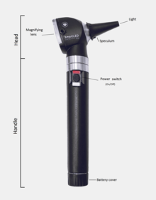
The head contains a light source and a magnifying lens, typically around 8 diopters (3× magnification),[citation needed] to help illuminate and enlarge ear structures.
It is important to brace the index or little finger of the hand holding the otoscope against the patient's head to avoid injuring the ear canal.
Most models also have an insertion point for a bulb that pushes air through the speculum (pneumatic otoscopy) for testing eardrum mobility.
The binocular view enables depth perception, which makes removal of earwax or other obstructing materials easier and less hazardous.
Studies have shown that reliance on a monocular otoscope to diagnose ear disease results in a more than 50% chance of misdiagnosis, as compared to binocular microscopic otoscopy.
By gently squeezing and releasing the bulb in rapid succession, the degree of eardrum mobility in response to positive and negative pressure can be observed.
The head is designed so that an airtight chamber is produced when a speculum is attached and fitted snugly into the patient's ear canal.