Panayiotopoulos syndrome
[1] An expert consensus has defined Panayiotopoulos syndrome as "a benign age-related focal seizure disorder occurring in early and mid-childhood.
Other autonomic manifestations include pallor (or, less often, flushing or cyanosis), mydriasis (or, less often, miosis), cardiorespiratory and thermoregulatory alterations, incontinence of urine and/or feces, hypersalivation, and modifications of intestinal motility.
[8] Characteristically, even after the most severe seizures and autonomic status epilepticus, the child is normal after a few hours of sleep, which is both diagnostic and reassuring.
Epileptic discharges in Panayiotopoulos syndrome, irrespective of their location at onset, activate emetic and autonomic centers prior to any other conventional neocortical seizure manifestations.
An explanation for this is that children are susceptible to autonomic disorders as illustrated by the cyclic vomiting syndrome, which is a nonepileptic condition specific to childhood.
[citation needed] Panayiotopoulos syndrome and all other benign childhood focal seizures, with rolandic epilepsy as their main representative, are probably linked due to a common, genetically determined, mild, and reversible functional derangement of the brain cortical maturational process that Panayiotopoulos proposed as "benign childhood seizure susceptibility syndrome".
A child with the distinctive clinical features of Panayiotopoulos syndrome, particularly ictus emeticus and lengthy seizures, may not need any investigations other than EEG.
The seizure may be very dramatic, with symptoms accumulating in succession, convulsions may occur and a child who becomes unresponsive and flaccid demands rigorous and experienced evaluation.
The most prominent acute disorders in the differential diagnosis include encephalitis or an encephalopathic state from causes such as infections, metabolic derangement (either inborn error or others such as hypoglycaemia), raised intracranial pressure and so forth.
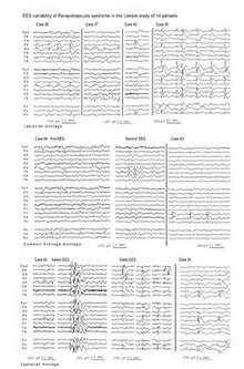
[citation needed] There are now significant reports of ictal EEGs in 20 cases, which objectively document the seizures of Panayiotopoulos syndrome and their variable localisation at onset.
The first clinical manifestation which appeared long (1–10 minutes) after the electrical onset, usually consisted of opening of the eyes as if the children were waking from sleep.
Of non-autonomic manifestations, deviation of eyes to the right or left occurred before or after vomiting without any apparent EEG localisation; it was present in seizures starting from the occipital or frontal regions.
[19][20] In a typical presentation of Panayiotopoulos syndrome, the child looks pale, vomits, and is fully conscious, able to speak, and understand but complains of "feeling sick."
This case illustrates autonomic status epilepticus with behavioral disturbances that would be difficult to attribute to seizure activity before the motor focal ictal events.
Within 5 minutes, he started banging his head on the wall and soon became unresponsive and floppy "like a rag doll," as well as incontinent of urine and feces with his eyes widely open and pupils markedly dilated.
This case also illustrates the features of syncope-like epileptic seizures together with other variable autonomic symptoms (emesis, respiratory abnormalities, pallor, mydriasis) in Panayiotopoulos syndrome.
A 5-year-old boy at age 13 months woke up vomiting profusely and then, while he was still in bed, became unresponsive and floppy with shallow breathing for 20 minutes.
A second episode occurred after 11 months, during sleep, and consisted of impairment of consciousness, hypotonia, deviation of the eyes to the right, hypersalivation, and right-sided clonic convulsions.
He was treated in a major teaching hospital with triple therapy for suspected encephalitis but in the third day after admission this was stopped and he was discharged home.
"[26] The distinctive clinical features particularly lengthy seizures and ictus emeticus means that the diagnosis of Panayiotopoulos syndrome is easy.
However, these are frequently mistaken as nonepileptic conditions such as acute encephalitis, syncope, migraine, cyclic vomiting syndrome, motion sickness, sleep disorder, or gastroenteritis.
The major problem is to recognize emetic and other autonomic manifestations as seizure events and not to dismiss them or erroneously to consider them as unrelated to the ictus and a feature of encephalitis, migraine, syncope or gastro-enteritis.
[28][29] Autonomic status epilepticus in the acute stage needs thorough evaluation for proper diagnosis and assessment of the neurologic/autonomic state of the child.
The traumatizing, sometimes long-lasting effect on parents is significant particularly because autonomic seizures may last for many hours compounded by physicians' uncertainty regarding diagnosis, management, and prognosis.
[38] However, though Panayiotopoulos syndrome is benign in terms of its evolution, autonomic seizures are potentially life-threatening in the rare context of cardiorespiratory arrest.
[citation needed] Chrysostomos (Tomis) P. Panayiotopoulos described this syndrome and autonomic status epilepticus particular to childhood through a 30-year prospective study that started in Greece in 1975.
[citation needed] In Panayiotopoulos' original study, ictal vomiting occurred in only 24 children out of 900 patients of all ages with epileptic seizures.
[citation needed] However, there was initial scepticism and resistance to these findings, including from influential epileptologists because as explained by Ferrie and Livingston:[44]"(a) ictal vomiting had been considered as extremely rare and hitherto had been mainly described in neurosurgical series of adult patients.
In children it was generally not considered as having an epileptic origin; (b) autonomic status epilepticus was not recognised as a diagnostic entity; the proposition that it might be a common occurrence in a benign seizure disorder challenged orthodox concepts of status epilepticus; (c) it implied that paediatricians had been failing to diagnose significant numbers of children with epilepsy, instead erroneously labeling then as having diverse non-epileptic disorders such as encephalitis, syncope, migraine, cyclic vomiting syndrome and gastroenteritis; (d) the characteristic EEG findings suggested alternative diagnoses.
[citation needed] The veracity of Panayiotopoulos's initial descriptions has, over the last two decades, been confirmed in large and long-term studies from Europe, Japan and South America.