Polyomaviridae
[11] Polyomaviruses are non-enveloped double-stranded DNA viruses with circular genomes of around 5000 base pairs.
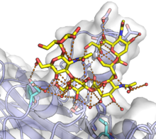
[12] The genome is packaged in a viral capsid of about 40-50 nanometers in diameter, which is icosahedral in shape (T=7 symmetry).
The early region usually codes for two proteins, the small and large tumor antigens, produced by alternative splicing.
The late region contains the three capsid structural proteins VP1, VP2, and VP3, produced by alternative translational start sites.
Cellular receptors for polyomaviruses are sialic acid residues of glycans, commonly gangliosides.
[16][18] However, in general virus-cell interactions are mediated by commonly occurring molecules on the cell surface, and therefore are likely not a major contributor to individual viruses' observed cell-type tropism.
[16] After binding to molecules on the cell surface, the virion is endocytosed and enters the endoplasmic reticulum - a behavior unique among known non-enveloped viruses[19] - where the viral capsid structure is likely to be disrupted by action of host cell disulfide isomerase enzymes.
[21] LT induces DNA replication from the viral genome's non-coding control region (NCCR), after which expression of the early mRNA is reduced and expression of the late mRNA, which encodes the viral capsid proteins, begins.
[16] Expression of the late genes results in accumulation of the viral capsid proteins in the host cell cytoplasm.
Capsid components enter the nucleus in order to encapsidate new viral genomic DNA.
[20] In some cases high levels of encapsidated virus result in cell lysis, releasing the virions.
[23] This is achieved by a two prong attack of inhibiting tumor suppressing genes p53 and members of the retinoblastoma (pRB) family,[24] and stimulating cell growth pathways by binding cellular DNA, ATPase-helicase, DNA polymerase α association, and binding of transcription preinitiation complex factors.
[citation needed] The small tumor antigen protein is also able to activate several cellular pathways that stimulate cell proliferation.
[29] Unlike for SV40, the MCV small T antigen directly transforms rodent cells in vitro.
Formerly, polyomaviruses and papillomaviruses, which share many structural features but have very different genomic organizations, were classified together in the now-obsolete family Papovaviridae.
[37] A subsequent proposed reclassification by the International Committee on Taxonomy of Viruses (ICTV) recommended dividing the family of Polyomaviridae into three genera:[38] The current ICTV classification system recognises six genera and 117 species, of which five could not be assigned a genus.
In the 1950s and early 1960s, well over 100 million people may have been exposed to SV40 due to previously undetected SV40 contamination of polio vaccine, prompting concern about the possibility that the virus might cause disease in humans.
[73] The reactivation of polyomavirus in the kidneys and urinary tract causes the shedding of infected cells, virions, and/or viral proteins in the urine.
[7][79][80][81] The JC virus offers a promising genetic marker for human evolution and migration.
[84] The causative agent was identified as a virus by Sarah Stewart and Bernice Eddy, after whom it was once called "SE polyoma".
[85][86][87] The term "polyoma" refers to the viruses' ability to produce multiple (poly-) tumors (-oma) under certain conditions.
The name has been criticized as a "meatless linguistic sandwich" ("meatless" because both morphemes in "polyoma" are affixes) giving little insight into the viruses' biology; in fact, subsequent research has found that most polyomaviruses rarely cause clinically significant disease in their host organisms under natural conditions.