Precession electron diffraction
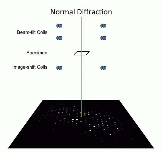
By rotating (precessing) a tilted incident electron beam around the central axis of the microscope, a PED pattern is formed by integration over a collection of diffraction conditions.
This produces a quasi-kinematical diffraction pattern that is more suitable as input into direct methods algorithms to determine the crystal structure of the sample.
These snapshots contain far fewer strongly excited reflections than a normal zone axis pattern and extend farther into reciprocal space.
[2] PED possesses many advantageous attributes that make it well suited to investigating crystal structures via direct methods approaches:[1] Precession electron diffraction is typically conducted using accelerating voltages between 100-400 kV.
[1] In choosing a precession rate, it is important to ensure that many revolutions of the beam occur over the relevant exposure time used to record the diffraction pattern.
Because PED takes the beam off of the optic axis by design, it accentuates the effect of the spherical aberrations within the probe forming lens.
50 nm is a general lower limit on probe size for standard TEMs operating at high precession angles (>30 mrad), but can be surpassed in Cs corrected instruments.
[5] If the precession angle is made too large, further complications due to the overlap of the ZOLZ and HOLZ reflections in the projected pattern can occur.
First, a Lorentz correction analogous to that used in x-ray diffraction can be applied to account for the fact that reflections are infrequently exactly at the Bragg condition over the course of a PED measurement.
This geometrical correction factor can be shown to assume the approximate form:[7] where g is the reciprocal space magnitude of the reflection in question and Ro is the radius of the Laue circle, usually taken to be equal to φ.
This form seeks to correct for both geometric and dynamical effects, but is still only an approximation that often fails to significantly improve the kinematic quality of the diffraction pattern (sometimes even worsening it).
More complete and accurate treatments of these theoretical correction factors have been shown to adjust measured intensities into better agreement with kinematical patterns.
The theory of precession electron diffraction is still an active area of research, and efforts to improve on the ability to correct measured intensities without a priori knowledge are ongoing.
This plug-in enables the widely used software package to collect precession electron diffraction patterns without additional modifications to the microscope.
Mapping the relative orientation of crystalline grains and/or phases helps understand material texture at the micro and nano scales.
In a transmission electron microscope, this is accomplished by recording a diffraction pattern at a large number of points (pixels) over a region of the crystalline specimen.
Because this process is highly automated, the quality of the recorded diffraction patterns is crucial to the software's ability to accurately compare and assign orientations to each pixel.
Tomography entails collecting a series of images (2-D projections) at various tilt angles and combining them to reconstruct the three dimensional structure of the specimen.
When probing a sample under diffraction conditions near a zone axis, as is often the case in EDS and EELS applications, channelling can have a large impact on the effective interaction of the incident electrons with specific ions in the crystal structure.
In practice, this can lead to erroneous measurements of composition that depend strongly on the orientation and thickness of the sample and the accelerating voltage.
Since PED entails an integration over incident directions of the electron probe, and generally does not include beams parallel to the zone axis, the detrimental channeling effects outlined above can be minimized, yielding far more accurate composition measurements in both techniques.