Pupillary light reflex
The pupillary light reflex (PLR) or photopupillary reflex is a reflex that controls the diameter of the pupil, in response to the intensity (luminance) of light that falls on the retinal ganglion cells of the retina in the back of the eye, thereby assisting in adaptation of vision to various levels of lightness/darkness.
When light is shone into only one eye and not the other, it is normal for both pupils to constrict simultaneously.
A consensual pupillary reflex is response of a pupil to light that enters the contralateral (opposite) eye.
Thus there are four types of pupillary light reflexes, based on this terminology of absolute laterality (left versus right) and relative laterality (same side versus opposite side, ipsilateral versus contralateral, direct versus consensual): The pupillary light reflex neural pathway on each side has an afferent limb and two efferent limbs.
Anatomically, the afferent limb consists of the retina, the optic nerve, and the pretectal nucleus in the midbrain, at level of superior colliculus.
Ganglion cells of the retina project fibers through the optic nerve to the ipsilateral pretectal nucleus.
The pretectal nucleus projects nerve fibers to the ipsilateral and contralateral Edinger-Westphal nuclei, which are also located in the midbrain.
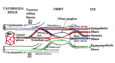
Postganglionic nerve fibers leave the ciliary ganglion to innervate the pupillary sphincter.
Sympathetic nervous system plays a role in dilating the pupils in low light conditions.
[4] The optic nerve, or more precisely, the photosensitive ganglion cells through the retinohypothalamic tract, is responsible for the afferent limb of the pupillary reflex; it senses the incoming light.
The oculomotor nerve is responsible for the efferent limb of the pupillary reflex; it drives the iris muscles that constrict the pupil.
Segments 7 and 8 each contains parasympathetic fibers that courses from the Edinger-Westphal nucleus, through the ciliary ganglion, along the oculomotor nerve (cranial nerve #3), to the ciliary sphincter, the muscular structure within the iris.
The diagram may assist in localizing lesion within the pupillary reflex system by process of elimination, using light reflex testing results obtained by clinical examination.
Pupillary light reflex provides a useful diagnostic tool for testing the integrity of the sensory and motor functions of the eye.
[1] Emergency physicians routinely test pupillary light reflex to assess brain stem function.
Abnormal pupillary reflex can be found in optic nerve injury, oculomotor nerve damage, brain stem lesion (including brain stem death), and depressant drugs, such as barbiturates.
Location of the lesion can be deduced as follows: The pupillary response to light is not purely reflexive, but is modulated by cognitive factors, such as attention, awareness, and the way visual input is interpreted.
[7][8] This shows that the pupillary light reflex is modulated by visual awareness.
[9][10][11] Moreover, the magnitude of the pupillary light reflex following a distracting probe is strongly correlated with the extent to which the probe captures visual attention and interferes with task performance.
[13][14] This shows that the pupillary light reflex is modulated by subjective (as opposed to objective) brightness.
is the pupillary latency, a time delay between the instant in which the light pulse reaches the retina and the beginning of iridal reaction due nerve transmission, neuro-muscular excitation and activation delays.
In order to improve the realism of the resulting simulations, the hippus effect can be approximated by adding small random variations to the environment light (in the range 0.05–0.3 Hz).