Respiratory system
The anatomy and physiology that make this happen varies greatly, depending on the size of the organism, the environment in which it lives and its evolutionary history.
[1] Gas exchange in the lungs occurs in millions of small air sacs; in mammals and reptiles, these are called alveoli, and in birds, they are known as atria.
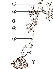
[2] These air sacs communicate with the external environment via a system of airways, or hollow tubes, of which the largest is the trachea, which branches in the middle of the chest into the two main bronchi.
In most fish, and a number of other aquatic animals (both vertebrates and invertebrates), the respiratory system consists of gills, which are either partially or completely external organs, bathed in the watery environment.
Gas exchange takes place in the gills which consist of thin or very flat filaments and lammellae which expose a very large surface area of highly vascularized tissue to the water.
Other animals, such as insects, have respiratory systems with very simple anatomical features, and in amphibians, even the skin plays a vital role in gas exchange.
[15][16][17]) It ends in the microscopic dead-end sacs called alveoli, which are always open, though the diameters of the various sections can be changed by the sympathetic and parasympathetic nervous systems.
[6] During heavy breathing (hyperpnea), as, for instance, during exercise, inhalation is brought about by a more powerful and greater excursion of the contracting diaphragm than at rest (Fig.
Seen from outside the body, the lifting of the clavicles during strenuous or labored inhalation is sometimes called clavicular breathing, seen especially during asthma attacks and in people with chronic obstructive pulmonary disease.
But now, the abdominal muscles, instead of remaining relaxed (as they do at rest), contract forcibly pulling the lower edges of the rib cage downwards (front and sides) (Fig. 8).
This not only drastically decreases the size of the rib cage, but also pushes the abdominal organs upwards against the diaphragm which consequently bulges deeply into the thorax (Fig. 8).
The abdominal muscles contract very powerfully, causing the pressure inside the abdomen and thorax to rise to extremely high levels.
The Valsalva maneuver can be carried out voluntarily but is more generally a reflex elicited when attempting to empty the abdomen during, for instance, difficult defecation, or during childbirth.
It is folded into about 300 million small air sacs called alveoli[23] (each between 75 and 300 μm in diameter) branching off from the respiratory bronchioles in the lungs, thus providing an extremely large surface area (approximately 145 m2) for gas exchange to occur.
[6] These areas form a series of neural pathways which receive information about the partial pressures of oxygen and carbon dioxide in the arterial blood.
[6] At sea level, under normal circumstances, the breathing rate and depth, is determined primarily by the arterial partial pressure of carbon dioxide rather than by the arterial partial pressure of oxygen, which is allowed to vary within a fairly wide range before the respiratory centers in the medulla oblongata and pons respond to it to change the rate and depth of breathing.
At altitude this causes the pulmonary arterial pressure to rise resulting in a much more even distribution of blood flow to the lungs than occurs at sea level.
[6] During coughing, contraction of the smooth muscle in the airway walls narrows the trachea by pulling the ends of the cartilage plates together and by pushing soft tissue into the lumen.
[36] Most of the respiratory system is lined with mucous membranes that contain mucosa-associated lymphoid tissue, which produces white blood cells such as lymphocytes.
[6] This causes them to have a greater surface tension-lowering effect when the alveoli are small than when they are large (as at the end of inhalation, when the surfactant molecules are more widely spaced).
[39] The lung vessels contain a fibrinolytic system that dissolves clots that may have arrived in the pulmonary circulation by embolism, often from the deep veins in the legs.
Horses are obligate nasal breathers which means that they are different from many other mammals because they do not have the option of breathing through their mouths and must take in air through their noses.
[41] This lack of a pleural space, along with an unusually thick diaphragm, are thought to be evolutionary adaptations allowing the elephant to remain underwater for long periods while breathing through its trunk which emerges as a snorkel.
[45] Inhalation and exhalation are brought about by alternately increasing and decreasing the volume of the entire thoraco-abdominal cavity (or coelom) using both their abdominal and costal muscles.
This pushes the sternal ribs, to which they are attached at almost right angles, downwards and forwards, taking the sternum (with its prominent keel) in the same direction (Fig. 17).
During exhalation the external oblique muscle which is attached to the sternum and vertebral ribs anteriorly, and to the pelvis (pubis and ilium in Fig.
Each pair of dorso-ventrobronchi is connected by a large number of parallel microscopic air capillaries (or parabronchi) where gas exchange occurs (Fig. 16).
The skin of these animals is highly vascularized and moist, with moisture maintained via secretion of mucus from specialised cells, and is involved in cutaneous respiration.
Back-flow into the gill chamber during the inhalatory phase is prevented by a membrane along the ventroposterior border of the operculum (diagram on the left in Fig.
As a result, labyrinth fish can survive for a short period of time out of water, as they can inhale the air around them, provided they stay moist.


Key:
1. skull ; 2. cervical vertebrae ; 3. furcula ; 4. coracoid ; 5. vertebral ribs ; 6. sternum and its keel ; 7. patella ; 8. tarsus ; 9. digits ; 10. tibia ( tibiotarsus ); 11. fibula ( tibiotarsus ); 12. femur ; 13. ischium ( innominate ); 14. pubis (innominate); 15. ilium (innominate); 16. caudal vertebrae ; 17. pygostyle ; 18. synsacrum ; 19. scapula ; 20. dorsal vertebrae ; 21. humerus ; 22. ulna ; 23. radius ; 24. carpus ( carpometacarpus ); 25. metacarpus ( carpometacarpus ); 26. digits ; 27. alula


