Roentgen (unit)
[3] It is named after the German physicist Wilhelm Röntgen, who discovered X-rays and was awarded the first Nobel Prize in Physics for the discovery.
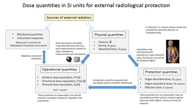
Additionally, a new quantity, kerma, was defined for air ionisation as the exposure for instrument calibration, and from this the absorbed dose can be calculated using known coefficients for specific target materials.
It was last defined by the U.S.'s National Institute of Standards and Technology (NIST) in 1998 as 2.58×10⁻⁴ C/kg, with a recommendation that the definition be given in every document where the roentgen is used.
In the meantime, the French Roentgen was given a different definition which amounted to 0.444 German R. In 1928, the International Congress of Radiology (ICR) defined the roentgen as "the quantity of X-radiation which, when the secondary electrons are fully utilised and the wall effect of the chamber is avoided, produce in 1 cc of atmospheric air at 0 °C and 76 cm of mercury pressure such a degree of conductivity that 1 esu of charge is measured at saturation current.
GOST standard 7623 defined it as "the physical dose of X-rays which produces charges each of one electrostatic unit in magnitude per cm3 of irradiated volume in air at 0 °C and normal atmospheric pressure when ionization is complete.
The International Commission on Radiation Units and Measurements (ICRU) took over the definition of the roentgen in 1950, defining it as "the quantity of X or γ-radiation such that the associated corpuscular emission per 0.001293 gram of air produces, in air, ions carrying 1 electrostatic unit of quantity of electricity of either sign.
"[15] The 3 MeV cap was no longer part of the definition, but the degraded usefulness of this unit at high beam energies was mentioned in the accompanying text.
The medical imaging community still has a need for ionization measurements, but they gradually converted to using C/kg as legacy equipment was replaced.
[17] In 1971 the European Economic Community, in Directive 71/354/EEC, catalogued the units of measure that could be used "for ... public health ...
The NIST brochures defined the roentgen as 2.58 × 10⁻⁴ C/kg, to be employed with exposures of x or γ radiation, but did not state the medium to be ionized.
In 1940, Louis Harold Gray, who had been studying the effect of neutron damage on human tissue, together with William Valentine Mayneord and the radiobiologist John Read, published a paper in which a unit of measure, dubbed the "gram roentgen" (symbol: gr) defined as "that amount of neutron radiation which produces an increment in energy in unit volume of tissue equal to the increment of energy produced in unit volume of water by one roentgen of radiation"[25] was proposed.
The definition of the roentgen had had the attraction of being relatively simple to define for photons in air, but the gray is independent of the primary ionizing radiation type, and can be used for both kerma and absorbed dose in a wide range of matter.
[29] When measuring absorbed dose in a human due to external exposure, the SI unit the gray, or the related non-SI rad are used.