Development of the human body
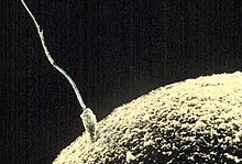
The process begins with fertilization, where an egg released from the ovary of a female is penetrated by a sperm cell from a male.
[2] The zygote contains a full complement of genetic material with all the biological characteristics of a single human being, and develops into the embryo.
Prior to implantation, the embryo remains in a protein shell, the zona pellucida, and undergoes a series of rapid mitotic cell divisions called cleavage.
[3] A week after fertilization the embryo still has not grown in size, but hatches from the zona pellucida and adheres to the lining of the mother's uterus.
[2] The genetic material of the sperm and egg then combine to form a single cell called a zygote and the germinal stage of prenatal development commences.
The entire process of embryonic development involves coordinated spatial and temporal changes in gene expression, cell growth and cellular differentiation.
A fetus is also characterized by the presence of all the major body organs, though they will not yet be fully developed and functional and some not yet situated in their final location.
The placenta connects the developing fetus to the uterine wall to allow nutrient uptake, thermo-regulation, waste elimination, and gas exchange via the mother's blood supply; to fight against internal infection; and to produce hormones which support pregnancy.
Placentas are a defining characteristic of placental mammals, but are also found in marsupials and some non-mammals with varying levels of development.
In response to the signals, the gonads produce hormones that stimulate libido and the growth, function, and transformation of the brain, bones, muscle, blood, skin, hair, breasts, and sex organs.
Until the maturation of their reproductive capabilities, the pre-pubertal physical differences between boys and girls are the external sex organs.
This begins in the third week of embryonic development, when the gastrula forms three distinct germ layers, the ectoderm, mesoderm and endoderm.
[22] During childhood, the bones undergo a complex process of elongation that occurs in a specific area called the epiphyseal growth plates (EGP).
These hormones promote the production of insulin-like growth factor-1 (IGF-1), which plays a key role in the formation of new bone cells.
[26] IGF-1 has six binding proteins (IGFBPs), exhibiting different effects on body tissues, where IGFBP-3 is most abundant in human circulation.
[27] IGF-1 initiates growth through differentiation and maturation of osteoblasts, and regulates release of GH from the pituitary through feedback mechanisms.
[25] At the same time inflammation and increased production of pro-inflammatory cytokines may cause GH resistance and a decrease in circulating IGF-1 and IGFBP-3 which in turn reduces endochondrial ossification and growth.
[31] In a large study based on 5 birth cohorts in Brazil, Guatemala, India, the Philippines and South Africa, faster linear growth at 0–2 years was associated with improvements in adult stature and school performance, but also an increased likelihood of overweight (mainly related to lean mass) and a slightly elevated blood pressure in young adulthood.