Sensory-motor coupling
"Neural responses at almost every stage of a sensorimotor pathway are modified at short and long timescales by biophysical and synaptic processes, recurrent and feedback connections, and learning, as well as many other internal and external variables".
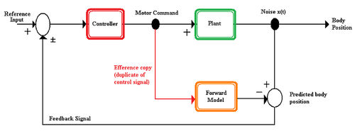
Flexible sensorimotor integration would allow an animal the ability to correct for errors and be useful in multiple situations.
[1][3] To produce the desired flexibility it's probable that nervous systems employ the use of internal models and efference copies.
The efference copy can be used by the nervous system to distinguish self-generated environmental changes, compare an expected response to what actually occurs in the environment, and to increase the rate at which a command can be issued by predicting an organism's state prior to receiving sensory input.
The assumption is that the nervous system has an internal representation of how a motor apparatus, the part of the body that will be moved, behaves in an environment.
The forward model takes the efference copy as an input and outputs the expected sensory changes.
[5] To reduce self-desensitization, the cricket's thoracic central pattern generator sends a corollary discharge, an efference copy that is used to inhibit an organism's response to self-generated stimuli, to the auditory system.
[1][5] The corollary discharge is used to inhibit the auditory system's response to the cricket's own song and prevent desensitization.
This prediction is used to check that the motor command will produce the goal sound so that corrections may be made.
was a man who suffered damage in his parietal and occipital lobes, areas of the brain related to processing visual information, due to a stroke.
[11] In fact, studies using external vibrations to create proprioceptive errors in movement show that Parkinson's patients perform better than healthy people.
In both quinolinic models and patients, it has been shown that people with Huntington's have abnormal sensory input.
The " various problems in integrating sensory information explain why patients with HD are unable to control voluntary movements accurately.
There are multiple pieces of evidence that indicate focal dystonia is related to improper linking or processing of afferent sensory information in the motor regions of the brain.
A sensory trick is the application of a stimulus to an area near to the location affected by dystonia that provides relief.
Research has shown that the motor cortex has increased excitability in RLS patients compared to healthy people.