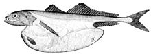
Fish anatomy
Low-frequency vibrations are detected by the lateral line system of sense organs that run along the length of the sides of fish, which responds to nearby movements and to changes in water pressure.
There are two different skeletal types: the exoskeleton, which is the stable outer shell of an organism, and the endoskeleton, which forms the support structure inside the body.
They function to move, support, and protect the various organs of the body, produce red and white blood cells and store minerals.
A similar arrangement was found in primitive tetrapods, but in the evolutionary line that led to reptiles, mammals and birds, the intercentrum became partially or wholly replaced by an enlarged pleurocentrum, which in turn became the bony vertebral body.
[11] In most ray-finned fishes, including all teleosts, these two structures are fused with and embedded within a solid piece of bone superficially resembling the vertebral body of mammals.
In living amphibians, there is simply a cylindrical piece of bone below the vertebral arch, with no trace of the separate elements present in the early tetrapods.
The upper tube is formed from the vertebral arches, but also includes additional cartilaginous structures filling in the gaps between the vertebrae, enclosing the spinal cord in an essentially continuous sheath.
The simpler structure is found in jawless fish, in which the cranium is represented by a trough-like basket of cartilaginous elements only partially enclosing the brain and associated with the capsules for the inner ears and the single nostril.
The cranium is a single structure forming a case around the brain, enclosing the lower surface and the sides, but always at least partially open at the top as a large fontanelle.
For suction feeding a system of connected four-bar linkages is responsible for the coordinated opening of the mouth and 3-D expansion of the buccal cavity.
[17] The ancestors of modern hagfish, thought to be protovertebrate,[18] were evidently pushed to very deep, dark waters, where they were less vulnerable to sighted predators and where it is advantageous to have a convex eyespot, which gathers more light than a flat or concave one.
It commonly has a number of pyloric caeca, small pouch-like structures along its length that help to increase the overall surface area of the organ for digesting food.
[32] In most herbivores the caecum receives partially digested food from the small intestine, and serves as a fermentation chamber to break down cellulose (such as grass or leaves) in the diet.
[40] In cartilaginous and bony fish it consists primarily of red pulp and is normally a somewhat elongated organ as it actually lies inside the serosal lining of the intestine.
Even in these animals, there is a diffuse layer of haematopoietic tissue within the gut wall, which has a similar structure to red pulp, and is presumed to be homologous to the spleen of higher vertebrates.
[51] The ossicles connect the gas bladder wall with Y-shaped lymph sinus that is next to the lymph-filled transverse canal joining the saccules of the right and left ears.
Gonadal sex is influenced by a number of factors, including cell-autonomous genetic mechanisms, endocrine, paracrine, behavioral, or environmental signals.
[53] Spermatogenesis in testes is a process in which spermatogonia differentiates into spermatocytes through mitosis and meiosis, which halves the number of chromosomes, creating haploid spermatids.
[41] Under a tough membranous shell, the tunica albuginea, the testis of some teleost fish, contains very fine coiled tubes called seminiferous tubules.
Fish can present cystic or semi-cystic spermatogenesis[definition needed] in relation to the release phase of germ cells in cysts to the lumen of the seminiferous tubules.
A slight swelling of the anterior end of the dorsal nerve cord is found in the lancelet, though it lacks the eyes and other complex sense organs comparable to those of vertebrates.
The front end of the nerve tube is expanded by a thickening of the walls and expansion of the central canal of spinal cord into three primary brain vesicles; the prosencephalon (forebrain), mesencephalon (midbrain) and rhombencephalon (hindbrain) then further differentiated in the various vertebrate groups.
[61] The resulting anatomy of the central nervous system, with a single, hollow ventral nerve cord topped by a series of (often paired) vesicles is unique to vertebrates.
One of the brain areas that receives primary input from the lateral line organ, the medial octavolateral nucleus, has a cerebellum-like structure, with granule cells and parallel fibers.
Each Mauthner cell has an axon that crosses over, innervating neurons at the same brain level and then travelling down through the spinal cord, making numerous connections as it goes.
The synapses generated by a Mauthner cell are so powerful that a single action potential gives rise to a major behavioral response: within milliseconds the fish curves its body into a C-shape, then straightens, thereby propelling itself rapidly forward.
They also possess an identifiable thymus and a well-developed spleen (their most important immune organ) where various lymphocytes, plasma cells and macrophages develop and are stored.
Chondrostean fish (sturgeons, paddlefish and bichirs) possess a major site for the production of granulocytes within a mass that is associated with the meninges, the membranes surrounding the central nervous system.
[73] In addition, teleost fish possess a thymus, spleen and scattered immune areas within mucosal tissues (e.g. in the skin, gills, gut and gonads).
Much like the mammalian immune system, teleost erythrocytes, neutrophils and granulocytes are believed to reside in the spleen whereas lymphocytes are the major cell type found in the thymus.